Anti-G6PD antibody
-
概述
- 产品描述Glucose-6-phosphate 1-dehydrogenase (G6PD) plays an important role in the pentose phosphate pathway. It is a member of the glucose-6-phosphate dehydrogenase family of proteins. G6PD is a ubiquitous enzyme that produces pentose sugars for nucleic acid synthesis, but is also involved in carbohydrate degradation, as it is one of the main producers of NADPH reducing power. G6PD has NADP as a co-factor and structural element. It can be found as a homodimer or homotetramer, and is primarily detected in lymphoblasts, granulocytes and sperm. Defects in G6PD can cause chronic non-spherocytic hemolytic anemia (CNSHA), especially in areas in which malaria is an epidemic. Individuals with a high level of G6PD-deficiency are at higher risk of acute hemolytic attacks.
- 产品名称Anti-G6PD antibody
- 分子量59 kDa
- 种属反应性Human,Mouse,Rat
- 验证应用WB,ICC,IHC-P
- 抗体类型兔多抗
- 免疫原Recombinant protein.
- 偶联Non-conjugated
-
性能
- 形态Liquid
- 浓度1 mg/mL.
- 存放说明Store at +4℃ after thawing. Aliquot store at -20℃ or -80℃. Avoid repeated freeze / thaw cycles.
- 存储缓冲液1*PBS (pH7.4), 0.2% BSA, 50% Glycerol. Preservative: 0.05% Sodium Azide.
- 亚型IgG
- 纯化方式Protein A purified.
- 亚细胞定位Cytoplasm. Nucleus. Membrane.
- 其它名称
- G6PD antibody
- G6PD_HUMAN antibody
- G6PD1 antibody
more
-
应用
WB: 1:1,000-1:2,000
ICC: 1:50-1:200
IHC-P: 1:50-1:200
-
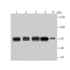
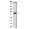
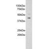
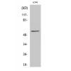
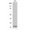
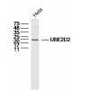
Fig1: Western blot analysis of G6PD on different lysates using anti-G6PD antibody at 1/1,000 dilution.
Positive control:
Lane 1: A549 Lane 2: Hela Lane 3: MCF-7 Lane 4: PC-12 Lane 5: Mouse spleen
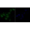
Fig2: ICC staining G6PD in A549 cells (green). The nuclear counter stain is DAPI (blue). Cells were fixed in paraformaldehyde, permeabilised with 0.25% Triton X100/PBS.
Fig3: ICC staining G6PD in Hela cells (green). The nuclear counter stain is DAPI (blue). Cells were fixed in paraformaldehyde, permeabilised with 0.25% Triton X100/PBS.
Fig4: ICC staining G6PD in MCF-7 cells (green). The nuclear counter stain is DAPI (blue). Cells were fixed in paraformaldehyde, permeabilised with 0.25% Triton X100/PBS.
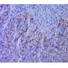
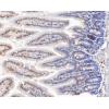
Fig5: Immunohistochemical analysis of paraffin-embedded human tonsil tissue using anti-G6PD antibody. Counter stained with hematoxylin.
Fig6: Immunohistochemical analysis of paraffin-embedded human colon cancer tissue using anti-G6PD antibody. Counter stained with hematoxylin.
Fig7: Immunohistochemical analysis of paraffin-embedded human spleen tissue using anti-G6PD antibody. Counter stained with hematoxylin.
Fig8: Immunohistochemical analysis of paraffin-embedded mouse testis tissue using anti-G6PD antibody. Counter stained with hematoxylin.
特别提示:本公司的所有产品仅可用于科研实验,严禁用于临床医疗及其他非科研用途!