Anti-CD319 antibody [5G5A4]
-
概述
- 产品描述Self-ligand receptor of the signaling lymphocytic activation molecule (SLAM) family. SLAM receptors triggered by homo- or heterotypic cell-cell interactions are modulating the activation and differentiation of a wide variety of immune cells and thus are involved in the regulation and interconnection of both innate and adaptive immune response. Activities are controlled by presence or absence of small cytoplasmic adapter proteins, SH2D1A/SAP and/or SH2D1B/EAT-2. Isoform 1 mediates NK cell activation through a SH2D1A-independent extracellular signal-regulated ERK-mediated pathway (PubMed:11698418). Positively regulates NK cell functions by a mechanism dependent on phosphorylated SH2D1B. Downstream signaling implicates PLCG1, PLCG2 and PI3K (PubMed:16339536). In addition to heterotypic NK cells-target cells interactions also homotypic interactions between NK cells may contribute to activation. However, in the absence of SH2D1B, inhibits NK cell function. Acts also inhibitory in T-cells (By similarity). May play a role in lymphocyte adhesion (PubMed:11802771). In LPS-activated monocytes negatively regulates production of proinflammatory cytokines (PubMed:23695528). Isoform 3 does not mediate any NK cell activation.
- 产品名称Anti-CD319 antibody [5G5A4]
- 分子量37.4kDa
- 种属反应性Human
- 验证应用WB,IHC-P,FC
- 抗体类型小鼠单抗
- 免疫原Purified recombinant fragment of human CD319 (AA: extra 23-226) expressed in E. Coli.
- 偶联Non-conjugated
-
性能
- 形态Liquid
- 浓度1 mg/mL
- 存放说明Store at +4℃ after thawing. Aliquot store at -20℃. Avoid repeated freeze / thaw cycles.
- 存储缓冲液1*PBS with 0.05% sodium azide.
- 亚型IgG1
- 纯化方式Protein G purified.
- 亚细胞定位Membrane.
- 其它名称
- 19A antibody
- 19A24 protein antibody
- CD 319 antibody
more
-
应用
WB: 1:500-1:2,000
IHC-P: 1:50-1:200
FC: 1:100-1:200
-
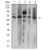
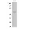
Fig1: Western blot analysis of CD319 against human CD319 (AA: extra 23-226) recombinant protein. Proteins were transferred to a PVDF membrane and blocked with 5% BSA in PBS for 1 hour at room temperature. The primary antibody was used in 5% BSA at room temperature for 2 hours. Goat Anti-Mouse IgG - HRP Secondary Antibody at 1:5,000 dilution was used for 1 hour at room temperature.
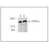
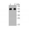
Fig2: Western blot analysis of CD319 against HEK293 (1) and CD319 (AA: extra 23-226)-hIgGFc transfected HEK293 (2) cell lysate. Proteins were transferred to a PVDF membrane and blocked with 5% BSA in PBS for 1 hour at room temperature. The primary antibody was used in 5% BSA at room temperature for 2 hours. Goat Anti-Mouse IgG - HRP Secondary Antibody at 1:5,000 dilution was used for 1 hour at room temperature.
Fig3: Immunohistochemical analysis of paraffin-embedded tonsil tissues using anti-CD319 antibody. The section was pre-treated using heat mediated antigen retrieval with Tris-EDTA buffer (pH 8.0) for 20 minutes. The tissues were blocked in 5% BSA for 30 minutes at room temperature, washed with ddH2O and PBS, and then probed with the primary antibody for 30 minutes at room temperature. The detection was performed using an HRP conjugated compact polymer system. DAB was used as the chromogen. Tissues were counterstained with hematoxylin and mounted with DPX.
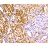
Fig4: Immunohistochemical analysis of paraffin-embedded rectum cancer tissues using anti-CD319 antibody. The section was pre-treated using heat mediated antigen retrieval with Tris-EDTA buffer (pH 8.0) for 20 minutes. The tissues were blocked in 5% BSA for 30 minutes at room temperature, washed with ddH2O and PBS, and then probed with the primary antibody for 30 minutes at room temperature. The detection was performed using an HRP conjugated compact polymer system. DAB was used as the chromogen. Tissues were counterstained with hematoxylin and mounted with DPX.
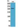
Fig5: Flow cytometric analysis of CD319 was done on Raji cells. The cells were fixed, permeabilized and stained with the primary antibody ) (green). After incubation of the primary antibody at room temperature for an hour, the cells were stained with a Alexa Fluor 488-conjugated goat anti-Mouse IgG Secondary antibody at 1/500 dilution for 30 minutes. Unlabelled sample was used as a control (cells without incubation with primary antibody; red).
特别提示:本公司的所有产品仅可用于科研实验,严禁用于临床医疗及其他非科研用途!

![Anti-CD319 antibody [5G5A4]](images/202012/goods_img/92566_G_1607495067653.jpg)