Anti-CD195 antibody [6G11D1]
-
概述
- 产品描述This gene encodes a member of the beta chemokine receptor family, which is predicted to be a seven transmembrane protein similar to G protein-coupled receptors. This protein is expressed by T cells and macrophages, and is known to be an important co-receptor for macrophage-tropic virus, including HIV, to enter host cells. Defective alleles of this gene have been associated with the HIV infection resistance. The ligands of this receptor include monocyte chemoattractant protein 2 (MCP-2), macrophage inflammatory protein 1 alpha (MIP-1 alpha), macrophage inflammatory protein 1 beta (MIP-1 beta) and regulated on activation normal T expressed and secreted protein (RANTES). Expression of this gene was also detected in a promyeloblastic cell line, suggesting that this protein may play a role in granulocyte lineage proliferation and differentiation. This gene is located at the chemokine receptor gene cluster region. An allelic polymorphism in this gene results in both functional and non-functional alleles; the reference genome represents the functional allele. Two transcript variants encoding the same protein have been found for this gene.
- 产品名称Anti-CD195 antibody [6G11D1]
- 分子量40.5kDa
- 种属反应性Human,Rat
- 验证应用WB,IHC-P,FC
- 抗体类型小鼠单抗
- 免疫原Purified recombinant fragment of human CD195 (AA: extra mix) expressed in E. Coli.
- 偶联Non-conjugated
-
性能
- 形态Liquid
- 浓度1 mg/mL
- 存放说明Store at +4℃ after thawing. Aliquot store at -20℃. Avoid repeated freeze / thaw cycles.
- 存储缓冲液1*PBS with 0.05% sodium azide.
- 亚型IgG1
- 纯化方式Protein G purified.
- 亚细胞定位Cell membrane.
- 其它名称
- AM4 7 antibody
- C C chemokine receptor type 5 antibody
- C C CKR 5 antibody
more
-
应用
WB: 1:500-1:2,000
IHC-P: 1:50-1:200
FC: 1:100-1:200
-
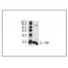
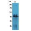
Fig1: Western blot analysis of CD195 against human CD195 (AA: extra mix) recombinant protein. Proteins were transferred to a PVDF membrane and blocked with 5% BSA in PBS for 1 hour at room temperature. The primary antibody was used in 5% BSA at room temperature for 2 hours. Goat Anti-Mouse IgG - HRP Secondary Antibody at 1:5,000 dilution was used for 1 hour at room temperature.
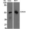
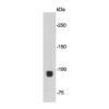
Fig2: Western blot analysis of CD195 against HEK293 (1) and CD195 (AA: extra mix)-hIgGFc transfected HEK293 (2) cell lysate. Proteins were transferred to a PVDF membrane and blocked with 5% BSA in PBS for 1 hour at room temperature. The primary antibody was used in 5% BSA at room temperature for 2 hours. Goat Anti-Mouse IgG - HRP Secondary Antibody at 1:5,000 dilution was used for 1 hour at room temperature.
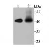
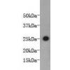
Fig3: Western blot analysis of CD195 against MOLT4 (1), L-02 (2), SPA-C-1 (3), A549 (4), and C6 (5) cell lysate. Proteins were transferred to a PVDF membrane and blocked with 5% BSA in PBS for 1 hour at room temperature. The primary antibody ( was used in 5% BSA at room temperature for 2 hours. Goat Anti-Mouse IgG - HRP Secondary Antibody at 1:5,000 dilution was used for 1 hour at room temperature.
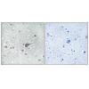
Fig4: Immunohistochemical analysis of paraffin-embedded cervical cancer tissues using anti-CD195 antibody. The section was pre-treated using heat mediated antigen retrieval with Tris-EDTA buffer (pH 8.0) for 20 minutes. The tissues were blocked in 5% BSA for 30 minutes at room temperature, washed with ddH2O and PBS, and then probed with the primary antibody (or 30 minutes at room temperature. The detection was performed using an HRP conjugated compact polymer system. DAB was used as the chromogen. Tissues were counterstained with hematoxylin and mounted with DPX.
Fig5: Immunohistochemical analysis of paraffin-embedded bladder cancer tissues using anti-CD195 antibody. The section was pre-treated using heat mediated antigen retrieval with Tris-EDTA buffer (pH 8.0) for 20 minutes. The tissues were blocked in 5% BSA for 30 minutes at room temperature, washed with ddH2O and PBS, and then probed with the primary antibody (E for 30 minutes at room temperature. The detection was performed using an HRP conjugated compact polymer system. DAB was used as the chromogen. Tissues were counterstained with hematoxylin and mounted with DPX.
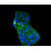
Fig6: Flow cytometric analysis of CD195 was done on K562 cells. The cells were fixed, permeabilized and stained with the primary antibody (green). After incubation of the primary antibody at room temperature for an hour, the cells were stained with a Alexa Fluor 488-conjugated goat anti-Mouse IgG Secondary antibody at 1/500 dilution for 30 minutes. Unlabelled sample was used as a control (cells without incubation with primary antibody; red).
特别提示:本公司的所有产品仅可用于科研实验,严禁用于临床医疗及其他非科研用途!

![Anti-CD195 antibody [6G11D1]](images/202012/goods_img/92017_G_1606897495055.jpg)