Anti-C-CBL antibody [C1-B12]
-
概述
- 产品名称Anti-C-CBL antibody [C1-B12]
- 分子量120 kDa
- 种属反应性Human,Mouse,Rat
- 验证应用WB,ICC,IHC-P,FC
- 抗体类型小鼠单抗
- 免疫原Recombinant protein
- 偶联Non-conjugated
-
性能
- 形态Liquid
- 浓度2 mg/mL.
- 存放说明Store at +4℃ after thawing. Aliquot store at -20℃ or -80℃. Avoid repeated freeze / thaw cycles.
- 存储缓冲液1*TBS (pH7.4), 1%BSA, Preservative: 0.05% Sodium Azide.
- 亚型IgG1
- 纯化方式Protein A purified.
- 亚细胞定位Cell membrane, Cytoplasm.
- 其它名称
- 4732447J05Rik antibody
- C CBL antibody
- Cas Br M (murine) ecotropic retroviral transforming sequence antibody
more
-
应用
WB: 1:500-1:1,000
ICC: 1:100-1:500
IHC-P: 1:50-1:200
FC: 1:100-1:200
-
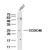
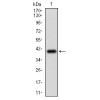
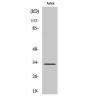
Fig1: Western blot analysis of CBL on human CBL recombinant protein using anti-CBL antibody at 1/1,000 dilution.
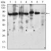
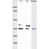
Fig2: Western blot analysis of CBL on different cell lysates using anti-CBL antibody at 1/1,000 dilution.
Positive control:
Lane 1: RAJI
Lane 2: RAW264.7
Lane 3: K562
Lane 4: SKBR-3
Lane 5: 3T3-L1
Lane 6: THP-1
Lane 7: PC-12
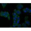
Fig3: ICC staining CBL (green) and actin filaments (red) in Hela cells. The nuclear counter stain is DAPI (blue). Cells were fixed in paraformaldehyde, permeabilised with 0.25% Triton X100/PBS.
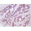
Fig4: Immunohistochemical analysis of paraffin-embedded ovarian cancer tissue using anti-CBL antibody. Counter stained with hematoxylin.
Fig5: Immunohistochemical analysis of paraffin-embedded bladder cancer tissue using anti-CBL antibody. Counter stained with hematoxylin.
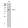
Fig6: Flow cytometric analysis of MCF-7 cells with CBL antibody at 1/100 dilution (blue) compared with an unlabelled control (cells without incubation with primary antibody; red).
特别提示:本公司的所有产品仅可用于科研实验,严禁用于临床医疗及其他非科研用途!


![Anti-C-CBL antibody [C1-B12]](images/202012/goods_img/92049_G_1606905156208.jpg)


