Anti-RAN antibody [A1-6D]
-
概述
- 产品描述The small Ras-related protein Ran, also called TC4, is a nuclear localized GTPase implicated in a diverse array of cellular processes including DNA replication, entry into and exit from mitosis and the transport of RNA and proteins through the nuclear pore complex. Like Ras, active Ran GTP and inactive Ran GDP levels are tightly regulated by guanine nucleotide exchange factors (GEFs) and GTPase activating proteins (GAPs). The abundant GEF, RCC1 (regulator of chromosome condensation 1), increases the rate at which Ran exchanges GDP for GTP. Ran GAP1 opposes the effects of RCC1 by increasing the rate at which Ran hydrolyzes GTP to GDP. A protein designated Ran BP1 has no intrinsic GAP activity, and functions as a GEF inhibitor deactivating RCC1 and thereby indirectly increasing the ratio of Ran GDP to Ran GTP. The Ran BP2 protein has been proposed as the Ran GTP docking site at the periphery of the nuclear pore complex.
- 产品名称Anti-RAN antibody [A1-6D]
- 分子量25 kDa
- 种属反应性Human,Mouse,Monkey,Rat
- 验证应用WB,IHC-P,ICC,FC
- 抗体类型小鼠单抗
- 免疫原Recombinant protein
- 偶联Non-conjugated
-
性能
- 形态Liquid
- 浓度2 mg/mL.
- 存放说明Store at +4℃ after thawing. Aliquot store at -20℃ or -80℃. Avoid repeated freeze / thaw cycles.
- 存储缓冲液1*TBS (pH7.4), 1%BSA, 40%Glycerol. Preservative: 0.05% Sodium Azide.
- 亚型IgG1
- 纯化方式Protein A purified.
- 亚细胞定位Nucleus. Cytoplasm. Melanosome.
- 其它名称
- Androgen receptor associated protein 24 antibody
- Androgen receptor-associated protein 24 antibody
- ARA 24 antibody
more
-
应用
WB: 1:500-1:2,000
ICC: 1:200-1:500
IHC-P: 1:200-1:500
FC: 1:100-1:200
-
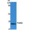
Fig1: Western blot analysis of RAN on human RAN recombinant protein using anti-RAN antibody at 1/1,000 dilution.
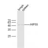
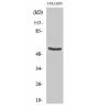
Fig2: Western blot analysis of RAN on HEK293 (1) and RAN-hIgGFc transfected HEK293 (2) cell lysate using anti-RAN antibody at 1/1,000 dilution.
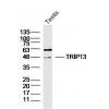
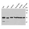
Fig3: Western blot analysis of RAN on different cell lysate using anti-RAN antibody at 1/1,000 dilution.
Positive control: Line1: Hela Line2: NIH/3T3 Line3: A431 Line4: C6 Line5: Jurkat Line6: Hela Line7: COS7 Line8: Jurkat
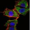
Fig4: ICC staining RAN (green) and Actin filaments (red) in GC-7901 cells. Cells were fixed in paraformaldehyde, permeabilised with 0.25% Triton X100/PBS.
Fig5: ICC staining RAN (green) and Actin filaments (red) in Hela cells. Cells were fixed in paraformaldehyde, permeabilised with 0.25% Triton X100/PBS.
Fig6: ICC staining RANRAN (green) and Actin filaments (red) in HepG2 cells. Cells were fixed in paraformaldehyde, permeabilised with 0.25% Triton X100/PBS.
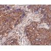
Fig7: Immunohistochemical analysis of paraffin-embedded human cervical cancer tissue using anti-RAN antibody. Counter stained with hematoxylin.
Fig8: Immunohistochemical analysis of paraffin-embedded human stomach cancer tissue using anti-RAN antibody. Counter stained with hematoxylin.
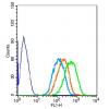
Fig9: Flow cytometric analysis of Hela cells with RAN antibody at 1/100 dilution (green) compared with an unlabelled control (cells without incubation with primary antibody; red).
特别提示:本公司的所有产品仅可用于科研实验,严禁用于临床医疗及其他非科研用途!


![Anti-RAN antibody [A1-6D]](images/202012/goods_img/92079_G_1606980655750.jpg)


