Anti-CBX2 antibody [C11-B10]
-
概述
- 产品描述Component of a Polycomb group (PcG) multiprotein PRC1-like complex, a complex class required to maintain the transcriptionally repressive state of many genes, including Hox genes, throughout development. PcG PRC1 complex acts via chromatin remodeling and modification of histones; it mediates monoubiquitination of histone H2A 'Lys-119', rendering chromatin heritably changed in its expressibility. Involved in sexual development, acting as activator of NR5A1 expression.
- 产品名称Anti-CBX2 antibody [C11-B10]
- 分子量56 kDa
- 种属反应性Human,Mouse
- 验证应用WB,ICC,IHC-P,FC
- 抗体类型小鼠单抗
- 免疫原Recombinant protein
- 偶联Non-conjugated
-
性能
- 形态Liquid
- 浓度2 mg/mL.
- 存放说明Store at +4℃ after thawing. Aliquot store at -20℃ or -80℃. Avoid repeated freeze / thaw cycles.
- 存储缓冲液1*TBS (pH7.4), 1%BSA, 40%Glycerol. Preservative: 0.05% Sodium Azide.
- 亚型IgG1
- 纯化方式Protein A purified.
- 亚细胞定位Nucleus
- 其它名称
- Cbx 2 antibody
- CBX2 antibody
- CBX2_HUMAN antibody
more
-
应用
WB: 1:500-1:2,000
ICC: 1:200-1:1,000
IHC-P: 1:200-1:500
FC: 1:50-1:200
-
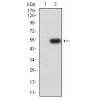
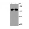
Fig1: Western blot analysis of CBX2 on human CBX2 recombinant protein using anti-CBX2 antibody at 1/1,000 dilution.
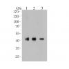
Fig2: Western blot analysis of CBX2 on HEK293 (1) and CBX2-hIgGFc transfected HEK293 (2) cell lysate using anti-CBX2 antibody at 1/1,000 dilution.
Fig3: Western blot analysis of CBX2 on different cell lysate using anti-CBX2 antibody at 1/1,000 dilution.
Positive control: Line1: HUVEC Line2: HEK293 Line3: Hela Line4: NIH/3T3 Line5: A431
Fig4: ICC staining CBX2 (green) and Actin filaments (red) in MCF-7 cells. The nuclear counter stain is DAPI (blue). Cells were fixed in paraformaldehyde, permeabilised with 0.25% Triton X100/PBS.
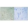
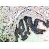
Fig5: Immunohistochemical analysis of paraffin-embedded human cervical cancer tissues using anti-CBX2 antibody. Counter stained with hematoxylin.
Fig6: Immunohistochemical analysis of paraffin-embedded human rectum cancer tissues using anti-CBX2 antibody. Counter stained with hematoxylin.
Fig7: Flow cytometric analysis of Hela cells with CBX2 antibody at 1/100 dilution (green) compared with an unlabelled control (cells without incubation with primary antibody; red).
特别提示:本公司的所有产品仅可用于科研实验,严禁用于临床医疗及其他非科研用途!

![Anti-CBX2 antibody [C11-B10]](images/202012/goods_img/92095_G_1606981976114.jpg)