Anti-PDE1B antibody [A3-C4]
-
概述
- 产品描述PDEs are key enzymes in signaling pathways that influence smooth muscle tone regulation. The PDE1 family are calmodulin-dependent (CaM-PDEs) that undergo stimulation through a calcium-calmodulin complex. Human PDE1B (PDE1B1) protein is present in neuronal cells of the cerebellum, hippocampus, and caudate, and lymphoblastoid lines, such as RPMI-8392 cells. PDE1B may participate in learning, memory, and regulation of phosphorylation of DARPP-32 in dopaminergic neurons. A splice variant known as PDE1B2 encodes a 516-amino acid protein and diverges from PDE1B1 by the replacement of the first 38 residues with an alternative 18 residues. The human PDE1B gene maps to chromosome 12q13, contains 13 exons, and encodes a 536 amino acid protein.
- 产品名称Anti-PDE1B antibody [A3-C4]
- 分子量61 kDa
- 种属反应性Human,Rat
- 验证应用WB,IHC-P,FC
- 抗体类型小鼠单抗
- 免疫原Recombinant protein
- 偶联Non-conjugated
-
性能
- 形态Liquid
- 浓度2 mg/mL.
- 存放说明Store at +4℃ after thawing. Aliquot store at -20℃ or -80℃. Avoid repeated freeze / thaw cycles.
- 存储缓冲液1*TBS (pH7.4), 1%BSA, 40%Glycerol. Preservative: 0.05% Sodium Azide.
- 亚型IgG1
- 纯化方式Protein A purified.
- 亚细胞定位Cytoplasm.
- 其它名称
- 5'-cyclic nucleotide phosphodiesterase 1B antibody
- 63 kDa Cam PDE antibody
- 63 kDa Cam-PDE antibody
more
-
应用
WB: 1:500-1:1,000
IHC-P: 1:50-1:200
FC: 1:100-1:200
-
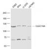
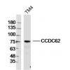
Fig1: Western blot analysis of PDE1B on human PDE1B recombinant protein using anti-PDE1B antibody at 1/1,000 dilution.
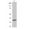
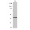
Fig2: Western blot analysis of PDE1B on HEK293 (1) and PDE1B-hIgGFc transfected HEK293 (2) cell lysate using anti-PDE1B antibody at 1/1,000 dilution.
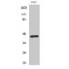
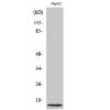
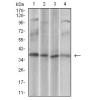
Fig3: Western blot analysis of PDE1B on PC-12 cell lysate using anti-PDE1B antibody at 1/1,000 dilution.
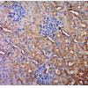
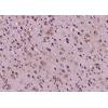
Fig4: Immunohistochemical analysis of paraffin-embedded human ovarian cancer tissue using anti-PDE1B antibody. Counter stained with hematoxylin.
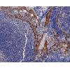
Fig5: Immunohistochemical analysis of paraffin-embedded human rectum cancer tissue using anti-PDE1B antibody. Counter stained with hematoxylin.
Fig6: Flow cytometric analysis of A549 cells with PDE1B antibody at 1/100 dilution (green) compared with an unlabelled control (cells without incubation with primary antibody; red).
特别提示:本公司的所有产品仅可用于科研实验,严禁用于临床医疗及其他非科研用途!

![Anti-PDE1B antibody [A3-C4]](images/202012/goods_img/92110_G_1606983527804.jpg)