Anti-PSMC3 antibody [C9-G10]
-
概述
- 产品描述In eukaryotic cells, selective breakdown of cellular proteins is ensured by their ubiquitination and subsequent degradation by the 26S Proteasome. The 26S Proteasome is a protease complex that selectively breaks down proteins that have been modified by polyubiquitin chains. It is made up of two multisubunit complexes: the 20S Proteasome chamber, which serves as the proteolytic core of the complex, and two 19S regulatory particles which recognize and unfold ubiquitinated proteins. PSMC3 (Proteasome 26S subunit ATPase 3), also known as TBP1 (Tat-binding protein 1), is a 439 amino acid member of the AAA ATPase family. Localized to both the nucleus and the cytoplasm, PSMC3 functions as a subunit of the 19S regulatory complex and is involved in regulating the substrate specificity of the 26S Proteasome. Additionally, PSMC3 interacts with the HIV protein HIV-1 Tat and, via this interaction, mediates the association of the viral protein with transcription complexes.
- 产品名称Anti-PSMC3 antibody [C9-G10]
- 分子量49 kDa
- 种属反应性Human,Monkey,Rat
- 验证应用WB,ICC,IHC-P,FC
- 抗体类型小鼠单抗
- 免疫原Recombinant protein
- 偶联Non-conjugated
-
性能
- 形态Liquid
- 浓度2 mg/mL.
- 存放说明Store at +4℃ after thawing. Aliquot store at -20℃ or -80℃. Avoid repeated freeze / thaw cycles.
- 存储缓冲液1*TBS (pH7.4), 1%BSA, 40%Glycerol. Preservative: 0.05% Sodium Azide.
- 亚型IgG1
- 纯化方式Protein A purified.
- 亚细胞定位Cytoplasm. Nucleus
- 其它名称
- 26S protease regulatory subunit 6A antibody
- 26S proteasome AAA-ATPase subunit RPT5 antibody
- Human immunodeficiency virus tat transactivator binding protein 1 antibody
more
-
应用
WB: 1:500-1:2,000
ICC: 1:50-1:200
IHC-P: 1:50-1:200
FC: 1:50-1:200
-
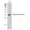
Fig1: Western blot analysis of PSMC3 on human PSMC3 recombinant protein using anti-PSMC3 antibody at 1/1,000 dilution.
Fig2: Western blot analysis of PSMC3 on HEK293 (1) and PSMC3-hIgGFc transfected HEK293 (2) cell lysate using anti-PSMC3 antibody at 1/1,000 dilution.
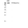
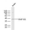
Fig3: Western blot analysis of PSMC3 on different cell lysate using anti-PSMC3 antibody at 1/1,000 dilution.
Positive control:
Lane 1: MCF-7
Lane 2: PC-3
Lane 3: T47D
Lane 4: SW620
Lane 5: COS7
Lane 6: C6
Lane 7: HELA
Lane 8: A431
Fig4: ICC staining PSMC3 (green) and Actin filaments (red) in MCF-7 cells. The nuclear counter stain is DAPI (blue). Cells were fixed in paraformaldehyde, permeabilised with 0.25% Triton X100/PBS.
Fig5: ICC staining PSMC3 (green) and Actin filaments (red) in SK-OV-3 cells. The nuclear counter stain is DAPI (blue). Cells were fixed in paraformaldehyde, permeabilised with 0.25% Triton X100/PBS.
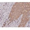
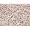
Fig6: Immunohistochemical analysis of paraffin-embedded human bladder cancer tissue using anti-PSMC3 antibody. Counter stained with hematoxylin.
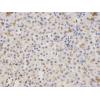
Fig7: Immunohistochemical analysis of paraffin-embedded human breast cancer tissue using anti-PSMC3 antibody. Counter stained with hematoxylin.
Fig8: Flow cytometric analysis of Hela cells with PSMC3 antibody at 1/100 dilution (green) compared with an unlabelled control (cells without incubation with primary antibody; red).
特别提示:本公司的所有产品仅可用于科研实验,严禁用于临床医疗及其他非科研用途!


![Anti-PSMC3 antibody [C9-G10]](images/202012/goods_img/92147_G_1606987059690.jpg)


