Anti-PLCG1 antibody [D11-C2]
-
概述
- 产品描述Phospholipase C-gamma 1 (PLC g1) is an isozyme of the phosphoinositide-specific PLC family, which occupies a central role in hormonal signal transduction pathways and is a substrate for the epidermal growth factor receptor tyrosine kinase. Following activation of TrkB, PLC g1 is phosphorylated on Tyrosine 783, Tyrosine 771 and Tyrosine 1253. Tyrosine 783 lies just downstream of the kinase domain in a relatively short sequence motif characteristic of the Trk family of protein-tyrosine kinase receptors. The sequence around Tyrosine 783 fits a consensus sequence for binding PLC g1. PLC g1 also forms a complex with TrkB consistent with the possibility that one of the TrkB autophosphorylation sites provides a binding site for the PLC g1 SH2 domains, as is the case for other receptor protein-tyrosine kinases.
- 产品名称Anti-PLCG1 antibody [D11-C2]
- 分子量148 kDa
- 种属反应性Human,Rat,Monkey
- 验证应用WB,ICC,IHC-P,FC
- 抗体类型小鼠单抗
- 免疫原Recombinant protein
- 偶联Non-conjugated
-
性能
- 形态Liquid
- 浓度2 mg/mL.
- 存放说明Store at +4℃ after thawing. Aliquot store at -20℃ or -80℃. Avoid repeated freeze / thaw cycles.
- 存储缓冲液1*TBS (pH7.4), 1%BSA, 40%Glycerol. Preservative: 0.05% Sodium Azide.
- 亚型IgG1
- 纯化方式Protein A purified.
- 亚细胞定位Cell projection, lamellipodium, Cell projection, ruffle
- 其它名称
- 1 phosphatidyl D myo inositol 4 5 bisphosphate antibody
- 1 phosphatidylinositol 4 5 bisphosphate phosphodiesterase gamma 1 antibody
- 1-phosphatidylinositol-4,5-bisphosphate phosphodiesterase gamma-1 antibody
more
-
应用
WB: 1:500-1:2,000
ICC: 1:50-1:200
IHC-P: 1:50-1:200
FC: 1:100-1:200
-
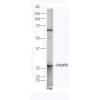
Fig1: Western blot analysis of PLCG1 on human PLCG1 recombinant protein using anti-PLCG1 antibody at 1/1,000 dilution.
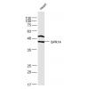
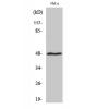
Fig2: Western blot analysis of PLCG1 on HEK293 (1) and PLCG1-hIgGFc transfected HEK293 (2) cell lysate using anti-PLCG1 antibody at 1/1,000 dilution.
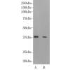
Fig3: f PLCG1 on different cell lysate using anti-PLCG1 antibody at 1/1,000 dilution.
Positive control: Line1: Hela Line2: A431 Line3: C6 Line4: NIH/3T3 Line5: COS7 Line6: HCT116
Fig4: ICC staining PLCG1 (green) and Actin filaments (red) in Hela cells. The nuclear counter stain is DAPI (blue). Cells were fixed in paraformaldehyde, permeabilised with 0.25% Triton X100/PBS.
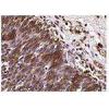
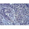
Fig5: Immunohistochemical analysis of paraffin-embedded human cervical cancer tissues using anti-PLCG1 antibody. Counter stained with hematoxylin.
Fig6: Immunohistochemical analysis of paraffin-embedded human bladder cancer tissues using anti-PLCG1 antibody. Counter stained with hematoxylin.
Fig7: Flow cytometric analysis of Jurkat cells with PLCG1 antibody at 1/100 dilution (green) compared with an unlabelled control (cells without incubation with primary antibody; red).
特别提示:本公司的所有产品仅可用于科研实验,严禁用于临床医疗及其他非科研用途!


![Anti-PLCG1 antibody [D11-C2]](images/202012/goods_img/92154_G_1606987564936.jpg)


