Anti-EZR antibody [F1-9A]
-
概述
- 产品描述Ezrin, Moesin and Radixin belong to a family of highly homologous actin-associated proteins that are localized just beneath the plasma membrane. The proteins are believed to be involved in the mediation of interactions between cytoskeletal and membrane proteins (2-5). Ezrin serves as a major cytoplasmic substrate of various protein-tyrosine kinases, including the epidermal growth factor receptor. Ezrin has also been identified as a cAMP-dependent protein kinase (A-kinase) anchoring protein and designated AKAP78. Moesin and Radixin share over 70% homology with Ezrin and are coexpressed within various cell types. Despite the high degree of homology, the three proteins exhibit a distinct receptor-specific pattern of phosphorylation.
- 产品名称Anti-EZR antibody [F1-9A]
- 分子量70 kDa
- 种属反应性Human,Mouse,Monkey
- 验证应用WB,ICC,FC
- 抗体类型小鼠单抗
- 免疫原Recombinant protein
- 偶联Non-conjugated
-
性能
- 形态Liquid
- 浓度2 mg/mL.
- 存放说明Store at +4℃ after thawing. Aliquot store at -20℃ or -80℃. Avoid repeated freeze / thaw cycles.
- 存储缓冲液1*TBS (pH7.4), 1%BSA, 40%Glycerol. Preservative: 0.05% Sodium Azide.
- 亚型IgG1
- 纯化方式Protein A purified.
- 亚细胞定位Apical cell membrane. Cell projection.
- 其它名称
- Villin 2 ezrin antibody
- CVIL antibody
- CVL antibody
more
-
应用
WB: 1:500-1:2,000
ICC: 1:50-1:200
FC: 1:50-1:100
-
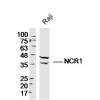
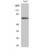
Fig1: Western blot analysis of EZR on human EZR recombinant protein using anti-EZR antibody at 1/1,000 dilution.
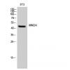
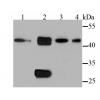
Fig2: Western blot analysis of EZR on HEK293 (1) and EZR-hIgGFc transfected HEK293 (2) cell lysate using anti-EZR antibody at 1/1,000 dilution.
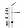
Fig3: Western blot analysis of EZR on different cell lysate using anti-EZR antibody at 1/1,000 dilution.
Positive control: Line1: MCF-7 Line2: Hela Line3: A431 Line4: Hek293 Line5: SK-N-SH Line6: Jurkat Line7: HepG2 Line8: NIH/3T3 Line9: Cos7
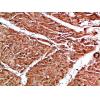
Fig4: ICC staining EZR (green) and Actin filaments (red) in A549 cells. The nuclear counter stain is DAPI (blue). Cells were fixed in paraformaldehyde, permeabilised with 0.25% Triton X100/PBS.
Fig5: Flow cytometric analysis of Hela cells with EZR antibody at 1/100 dilution (green) compared with an unlabelled control (cells without incubation with primary antibody; red).
Fig6: Flow cytometric analysis of MCF-7 cells with EZR antibody at 1/100 dilution (green) compared with an unlabelled control (cells without incubation with primary antibody; red).
特别提示:本公司的所有产品仅可用于科研实验,严禁用于临床医疗及其他非科研用途!

![Anti-EZR antibody [F1-9A]](images/202012/goods_img/92157_G_1606987737743.jpg)