Anti-CD31/PECAM-1 antibody [7-A1]
-
概述
- 产品描述Cell adhesion molecules are a family of closely related cell surface glycoproteins involved in cell-cell interactions during growth and are thought to play an important role in embryogenesis and development. Neuronal cell adhesion molecule (NCAM) expression is observed in a variety of human tumors including neuroblastomas, rhabdomyosarcomas, Wilms’ tumors, Ewing’s sarcomas and some primitive myeloid malignancies. The intracellular adhesion molecule-1 (ICAM-1), also referred to as CD54, is an integral membrane protein of the immunoglobulin superfamily. PECAM-1 (platelet/endothelial cell adhesion molecule-1), also referred to as CD31, is a glycoprotein expressed on the cell surfaces of monocytes, neutrophils, platelets and a subpopulation of T cells. VCAM-1 (vascular cell adhesion molecule-1) was first identified as an adhesion molecule induced on human endothelial cells by inflammatory cytokines such as IL-1, tumor necrosis factor (TNF) and lipopolysaccharide (LPS). The KALIG gene encodes a nerve cell adhesion molecule (NCAM)-like protein and is deleted in 66% of patients with Kallmann’s syndrome, anosmia with secondary hypogonadism.
- 产品名称Anti-CD31/PECAM-1 antibody [7-A1]
- 分子量82kDa(Observed: 130 kDa)
- 种属反应性Human,Mouse
- 验证应用WB,ICC/IF,IHC-P,FC
- 抗体类型小鼠单抗
- 免疫原Synthetic peptide (KLH-coupled) within C-terminal residues of human CD31.
- 偶联Non-conjugated
-
性能
- 形态Liquid
- 浓度2 mg/mL.
- 存放说明Store at +4℃ after thawing. Aliquot store at -20℃. Avoid repeated freeze / thaw cycles.
- 存储缓冲液1*PBS (pH7.4), 0.2% BSA, 40% Glycerol. Preservative: 0.05% Sodium Azide.
- 亚型IgG2a
- 纯化方式Peptide affinity purified
- 亚细胞定位Cell junction. Cell membrane. Membrane.
- 其它名称
- Adhesion molecule antibody
- CD31 antibody
- CD31 antigen antibody
more
-
应用
WB: 1:1,000-1:5,000
ICC/IF: 1:50-1:200
IHC-P: 1:2,000-1:5,000
FC:1:50-1:100
-
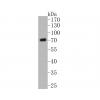
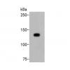
Fig1:
Western blot analysis of CD31 on THP-1 cells lysates using anti-CD31 antibody at 1/1,000 dilution.
Fig2: ICC staining CD31 in HUVEC cells (green). The nuclear counter stain is DAPI (blue). Cells were fixed in paraformaldehyde, permeabilised with 0.25% Triton X100/PBS.
Fig3: ICC staining CD31 in PMVEC cells (green). The nuclear counter stain is DAPI (blue). Cells were fixed in paraformaldehyde, permeabilised with 0.25% Triton X100/PBS.
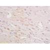
Fig4: Immunohistochemical analysis of paraffin-embedded human tonsil tissue using anti-CD31 antibody. Counter stained with hematoxylin.
Fig5: Immunohistochemical analysis of paraffin-embedded human kidney tissue using anti-CD31 antibody. Counter stained with hematoxylin.
Fig6: Immunohistochemical analysis of paraffin-embedded human uterus tissue using anti-CD31 antibody. Counter stained with hematoxylin.
Fig7: Immunohistochemical analysis of paraffin-embedded mouse kidney tissue using anti-CD31 antibody. Counter stained with hematoxylin.
Fig8: Immunohistochemical analysis of paraffin-embedded mouse uterus muscle tissue using anti-CD31 antibody. Counter stained with hematoxylin.
Fig9: Flow cytometric analysis of Jurkat cells with CD31 antibody at 1/100 dilution (red) compared with an unlabelled control (cells without incubation with primary antibody; black). Alexa Fluor 488-conjugated Goat anti mouse IgG was used as the secondary
特别提示:本公司的所有产品仅可用于科研实验,严禁用于临床医疗及其他非科研用途!


![Anti-CD31/PECAM-1 antibody [7-A1]](images/202012/goods_img/92202_G_1607071653739.jpg)


