Anti-DAP Kinase 1 antibody [12B2]
-
概述
- 产品描述Calcium/calmodulin-dependent serine/threonine kinase involved in multiple cellular signaling pathways that trigger cell survival, apoptosis, and autophagy. Regulates both type I apoptotic and type II autophagic cell deaths signal, depending on the cellular setting. The former is caspase-dependent, while the latter is caspase-independent and is characterized by the accumulation of autophagic vesicles. Phosphorylates PIN1 resulting in inhibition of its catalytic activity, nuclear localization, and cellular function. Phosphorylates TPM1, enhancing stress fiber formation in endothelial cells. Phosphorylates STX1A and significantly decreases its binding to STXBP1. Phosphorylates PRKD1 and regulates JNK signaling by binding and activating PRKD1 under oxidative stress. Phosphorylates BECN1, reducing its interaction with BCL2 and BCL2L1 and promoting the induction of autophagy. Phosphorylates TSC2, disrupting the TSC1-TSC2 complex and stimulating mTORC1 activity in a growth factor-dependent pathway. Phosphorylates RPS6, MYL9 and DAPK3. Acts as a signaling amplifier of NMDA receptors at extrasynaptic sites for mediating brain damage in stroke. Cerebral ischemia recruits DAPK1 into the NMDA receptor complex and it phosphorylates GRINB at Ser-1303 inducing injurious Ca2+ influx through NMDA receptor channels, resulting in an irreversible neuronal death. Required together with DAPK3 for phosphorylation of RPL13A upon interferon-gamma activation which is causing RPL13A involvement in transcript-selective translation inhibition.
- 产品名称Anti-DAP Kinase 1 antibody [12B2]
- 分子量160 kDa
- 种属反应性Human,Mouse,Rat
- 验证应用WB,IHC-P
- 抗体类型小鼠单抗
- 免疫原Recombinant protein within Human DAP Kinase 1 aa 450-670.
- 偶联Non-conjugated
-
性能
- 形态Liquid
- 浓度2 mg/mL.
- 存放说明Store at +4℃ after thawing. Aliquot store at -20℃. Avoid repeated freeze / thaw cycles.
- 存储缓冲液1*PBS (pH7.4), 0.2% BSA, 50% Glycerol. Preservative: 0.05% Sodium Azide.
- 亚型IgG1
- 纯化方式Protein affinity purified.
- 亚细胞定位Cytoplasm, Cytoskeleton.
- 其它名称
- DAK1 antibody
- DAP K1 antibody
- DAP kinase 1 antibody
more
-
应用
WB:1:500
IHC-P:1:50-1:200
-
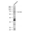
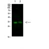
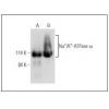
Fig1: Western blot analysis of DAP Kinase 1 on rat brain tissue lysate. Proteins were transferred to a PVDF membrane and blocked with 5% BSA in PBS for 1 hour at room temperature. The primary antibody was used at a 1:500 dilution in 5% BSA at room temperature for 2 hours. Goat Anti-Mouse IgG - HRP Secondary Antibody (HA1006) at 1:5,000 dilution was used for 1 hour at room temperature.
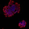
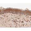
Fig2: Immunohistochemical analysis of paraffin-embedded rat lung tissue using anti-DAP Kinase 1 antibody. The section was pre-treated using heat mediated antigen retrieval with Tris-EDTA buffer (pH 8.0-8.4) for 20 minutes.The tissues were blocked in 5% BSA for 30 minutes at room temperature, washed with ddH2O and PBS, and then probed with the antibody at 1/50 dilution, for 30 minutes at room temperature and detected using an HRP conjugated compact polymer system. DAB was used as the chrogen. Counter stained with hematoxylin and mounted with DPX.
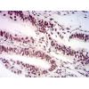
Fig3: Immunohistochemical analysis of paraffin-embedded human lung cancer tissue using anti-DAP Kinase 1 antibody. The section was pre-treated using heat mediated antigen retrieval with Tris-EDTA buffer (pH 8.0-8.4) for 20 minutes.The tissues were blocked in 5% BSA for 30 minutes at room temperature, washed with ddH2O and PBS, and then probed with the antibody at 1/200 dilution, for 30 minutes at room temperature and detected using an HRP conjugated compact polymer system. DAB was used as the chrogen. Counter stained with hematoxylin and mounted with DPX.
Fig4: Immunohistochemical analysis of paraffin-embedded human skin tissue using anti-DAP Kinase 1 antibody. The section was pre-treated using heat mediated antigen retrieval with Tris-EDTA buffer (pH 8.0-8.4) for 20 minutes.The tissues were blocked in 5% BSA for 30 minutes at room temperature, washed with ddH2O and PBS, and then probed with the antibody at 1/200 dilution, for 30 minutes at room temperature and detected using an HRP conjugated compact polymer system. DAB was used as the chrogen. Counter stained with hematoxylin and mounted with DPX.
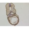
Fig5: Immunohistochemical analysis of paraffin-embedded mouse liver tissue using anti-DAP Kinase 1 antibody. The section was pre-treated using heat mediated antigen retrieval with Tris-EDTA buffer (pH 8.0-8.4) for 20 minutes.The tissues were blocked in 5% BSA for 30 minutes at room temperature, washed with ddH2O and PBS, and then probed with the antibody at 1/50 dilution, for 30 minutes at room temperature and detected using an HRP conjugated compact polymer system. DAB was used as the chrogen. Counter stained with hematoxylin and mounted with DPX.
特别提示:本公司的所有产品仅可用于科研实验,严禁用于临床医疗及其他非科研用途!


![Anti-DAP Kinase 1 antibody [12B2]](images/202012/goods_img/92230_G_1607167675704.jpg)


