Anti-TNFR1 antibody [2G2]
-
概述
- 产品描述Tumor necrosis factor (TNF) is a pleiotropic cytokine whose function is mediated through two distinct cell surface receptors. These receptors, designated TNF-R1 and TNF-R2 are expressed on most cell types. The majority of TNF functions are primarily mediated through TNF-R1, while signaling through TNF-R2 occurs less extensively and is confined to cells of the immune system. Both of these proteins belong to the growing TNF and nerve growth factor (NGF) receptor superfamily, which includes FAS, CD30, CD27 and CD40. The members of this superfamily are type I membrane proteins that share sequence homology confined to the extracellular region. TNF-R1 shares a motif coined the “death domain” with FAS and three structurally unrelated signaling proteins, TRADD, FADD and RIP. This “death domain” is required for transduction of the apoptotic signal.
- 产品名称Anti-TNFR1 antibody [2G2]
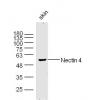
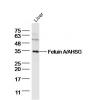
- 分子量55 kDa
- 种属反应性Human,Mouse,Rat
- 验证应用WB,IP,IF,IHC-P
- 抗体类型小鼠单抗
- 免疫原Amino acids 30-301 mapping within the extracellular domain of TNF-R1 of human origin.
- 偶联Non-conjugated
-
性能
- 形态Liquid
- 浓度2 mg/mL.
- 存放说明Store at +4℃
- 存储缓冲液1*TBS (pH7.4), 1%BSA, 40%Glycerol. Preservative: 0.05% Sodium Azide.
- 亚型IgG
- 纯化方式Immunogen affinity purified
- 亚细胞定位Secreted, Golgi apparatus membrane Cell membrane
- 其它名称
- CD120a antibody
- FPF antibody
- MGC19588 antibody
more
-
应用
WB: 1:100-1:1,000
IP: 1-2 μg per 100-500 μg of total protein(1 ml of cell lysate)
IF: 1:50-1:500
IHC-P: 1:50-1:500
-
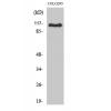
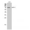
Fig1: Western blot analysis of TNF-R1 expression in MCF7 (A), HeLa (B) and U-937 (C) whole cell lysates.
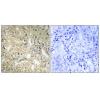
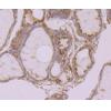
Fig2: Immunoperoxidase staining of formalin fixed, paraffin-embedded human small intestine tissue showing cytoplasmic staining of glandular cells.
特别提示:本公司的所有产品仅可用于科研实验,严禁用于临床医疗及其他非科研用途!


![Anti-TNFR1 antibody [2G2]](images/202012/goods_img/92310_G_1607243057260.jpg)


