Anti-Cytochrome C antibody [10-E11-G2]
-
概述
- 产品描述Cytochrome c is a well characterized mobile electron transport protein that is essential to energy conversion in all aerobic organisms. In mammalian cells, this highly conserved protein is normally localized to the mitochondrial intermembrane space. More recent studies have identifed cytosolic cytochrome c as a factor necessary for activation of apoptosis. During apoptosis, cytochrome c is translocated from the mitochondrial membrane to the cytosol, where it is required for activation of caspase-3 (CPP32). Overexpression of Bcl-2 has been shown to prevent the translocation of cytochrome c, thereby blocking the apoptotic process. Overexpression of Bax has been shown to induce the release of cytochrome c and to induce cell death. The release of cytochrome c from the mitochondria is thought to trigger an apoptotic cascade, whereby Apaf-1 binds to Apaf-3 (caspase-9) in a cytochrome c-dependent manner, leading to caspase-9 cleavage of caspase-3.
- 产品名称Anti-Cytochrome C antibody [10-E11-G2]
- 分子量12 kDa
- 种属反应性Human,Mouse,Rat
- 验证应用WB,IHC-P,ICC
- 抗体类型小鼠单抗
- 免疫原Peptide
- 偶联Non-conjugated
-
性能
- 形态Liquid
- 浓度2 mg/mL.
- 存放说明Store at +4℃ after thawing. Aliquot store at -20℃ or -80℃. Avoid repeated freeze / thaw cycles.
- 存储缓冲液1*PBS (pH7.4), 0.2% BSA, 50% Glycerol. Preservative: 0.05% Sodium Azide.
- 亚型IgG1
- 纯化方式Peptide affinity purified
- 亚细胞定位Mitochondrion.
- 其它名称
- CYC antibody
- CYC_HUMAN antibody
- CYCS antibody
more
-
应用
WB: 1:200-1:500
ICC: 1:50-1:200
IHC-P: 1:50-1:200
-
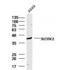
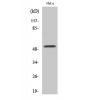
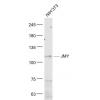
Fig1: Western blot analysis of Cytochrome C on mouse heart tissue lysate using anti-Cytochrome C antibody at 1/200 dilution.
Fig2: ICC staining Cytochrome C (red) in Hela cells. The nuclear counter stain is DAPI (blue). Cells were fixed in paraformaldehyde, permeabilised with 0.25% Triton X100/PBS.
Fig3: ICC staining Cytochrome C (red) in HepG2 cells. The nuclear counter stain is DAPI (blue). Cells were fixed in paraformaldehyde, permeabilised with 0.25% Triton X100/PBS.
Fig4: ICC staining Cytochrome C (red) in MCF-7 cells. The nuclear counter stain is DAPI (blue). Cells were fixed in paraformaldehyde, permeabilised with 0.25% Triton X100/PBS.
Fig5: Immunohistochemical analysis of paraffin-embedded human liver tissue using anti-Cytochrome C antibody. Counter stained with hematoxylin.
Fig6: Immunohistochemical analysis of paraffin-embedded human spleen tissue using anti-Cytochrome C antibody. Counter stained with hematoxylin.
Fig7: Immunohistochemical analysis of paraffin-embedded human kidney tissue using anti-Cytochrome C antibody. Counter stained with hematoxylin.
Fig8: Immunohistochemical analysis of paraffin-embedded mouse heart tissue using anti-Cytochrome C antibody. Counter stained with hematoxylin.
特别提示:本公司的所有产品仅可用于科研实验,严禁用于临床医疗及其他非科研用途!


![Anti-Cytochrome C antibody [10-E11-G2]](images/202012/goods_img/92347_G_1607249118128.jpg)


