Anti-PRDX6 antibody [7G1]
-
概述
- 产品描述The peroxiredoxin (PRX) family comprises six antioxidant proteins, PRX I, II, III, IV, V and VI, which protect cells from reactive oxygen species (ROS) by preventing the metal-catalyzed oxidation of enzymes. The PRX proteins primarily utilize thioredoxin as the electron donor for antioxidation, although they are fairly promiscuous with regard to the hydroperoxide substrate. In addition to protection from ROS, peroxiredoxins are also involved in cell proliferation, differentiation and gene expression. PRX I, II, IV and VI show diffuse cytoplasmic localization, while PRX III and V exhibit distinct mitochondrial localization.PRX VI, a 1-Cys peroxiredoxin (also known as antioxidant protein 2 or AOP2), is highly expressed in most tissues, particularly in epithelial cells. Localized to the cell cytosol, PRX VI functions independently of other peroxiredoxins and antioxidant proteins, specializing in antioxidant defense, lung phospholipid metabolism and protection of keratinocytes from cell death induced by reactive oxygen species.
- 产品名称Anti-PRDX6 antibody [7G1]
- 分子量25 kDa
- 种属反应性Human
- 验证应用WB,IHC-P,ICC,FC
- 抗体类型小鼠单抗
- 免疫原Recombinant protein.
- 偶联Non-conjugated
-
性能
- 形态Liquid
- 浓度2 mg/mL.
- 存放说明Store at +4℃ after thawing. Aliquot store at -20℃. Avoid repeated freeze / thaw cycles.
- 存储缓冲液1*PBS (pH7.4), 0.2% BSA, 50% Glycerol. Preservative: 0.05% Sodium Azide.
- 亚型IgG1
- 纯化方式Protein G purified.
- 亚细胞定位Cytoplasm. Lysosome.
- 其它名称
-
- 1 Cys antibody
- 1 Cys peroxiredoxin antibody
- 1 Cys PRX antibody
more
-
应用
WB: 1:500
ICC: 1:50-1:200
IHC-P: 1:100-1:400
FC: 1:50-1:100
-
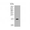
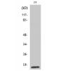
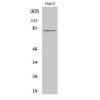
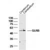
Fig1: Western blot analysis of PRDX6 on PC-3M (1) and K562 (2) using anti-PRDX6 antibody at 1/500 dilution.
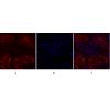
Fig2: ICC staining PRDX6 (green) in A431 cells. The nuclear counter stain is DAPI (blue). Cells were fixed in paraformaldehyde, permeabilised with 0.25% Triton X100/PBS.
Fig3: ICC staining PRDX6 (green) in PC-3M cells. The nuclear counter stain is DAPI (blue). Cells were fixed in paraformaldehyde, permeabilised with 0.25% Triton X100/PBS.
Fig4: ICC staining PRDX6 (green) in SiHa cells. The nuclear counter stain is DAPI (blue). Cells were fixed in paraformaldehyde, permeabilised with 0.25% Triton X100/PBS.
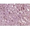
Fig5: Immunohistochemical analysis of paraffin-embedded human liver tissue using anti-PRDX6 antibody. Counter stained with hematoxylin.
Fig6: Immunohistochemical analysis of paraffin-embedded human colon cancer tissue using anti-PRDX6 antibody. Counter stained with hematoxylin.
Fig7: Immunohistochemical analysis of paraffin-embedded human kidney tissue using anti-PRDX6 antibody. Counter stained with hematoxylin.
Fig8: Immunohistochemical analysis of paraffin-embedded human placenta tissue using anti-PRDX6 antibody. Counter stained with hematoxylin.
Fig9: Flow cytometric analysis of PC-3M cells with PRDX6 antibody at 1/100 dilution (red) compared with an unlabelled control (cells without incubation with primary antibody; black). Alexa Fluor 488-conjugated goat anti-mouse IgG was used as the secondary
特别提示:本公司的所有产品仅可用于科研实验,严禁用于临床医疗及其他非科研用途!

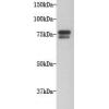
![Anti-PRDX6 antibody [7G1]](images/202012/goods_img/92540_G_1607424448366.jpg)