Anti-CD1C antibody [3G1B3]
-
概述
- 产品描述This gene encodes a member of the CD1 family of transmembrane glycoproteins, which are structurally related to the major histocompatibility complex (MHC) proteins and form heterodimers with beta-2-microglobulin. The CD1 proteins mediate the presentation of primarily lipid and glycolipid antigens of self or microbial origin to T cells. The human genome contains five CD1 family genes organized in a cluster on chromosome 1. The CD1 family members are thought to differ in their cellular localization and specificity for particular lipid ligands. The protein encoded by this gene is broadly distributed throughout the endocytic system via a tyrosine-based motif in the cytoplasmic tail. Alternatively spliced transcript variants of this gene have been observed, but their full-length nature is not known.
- 产品名称Anti-CD1C antibody [3G1B3]
- 分子量37.7kDa
- 种属反应性Human
- 验证应用WB,IHC-P,FC
- 抗体类型小鼠单抗
- 免疫原Purified recombinant fragment of human CD1C (AA: extra 18-302) expressed in E. Coli.
- 偶联Non-conjugated
-
性能
- 形态Liquid
- 浓度1 mg/mL
- 存放说明Store at +4℃ after thawing. Aliquot store at -20℃. Avoid repeated freeze / thaw cycles.
- 存储缓冲液1*PBS with 0.05% sodium azide.
- 亚型IgG2b
- 纯化方式Protein G purified.
- 亚细胞定位Cell membrane. Endosome membrane. Subject to intracellular trafficking between the cell membrane and endosomes.
- 其它名称
- BDCA1 antibody
- CD1 antibody
- CD1A antibody
more
-
应用
WB: 1:500-1:2,000
IHC-P: 1:50-1:200
FC: 1:100-1:200
-
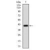
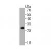
Fig1: Western blot analysis of CD1C against human CD1C (AA: extra 18-302) recombinant protein. Proteins were transferred to a PVDF membrane and blocked with 5% BSA in PBS for 1 hour at room temperature. The primary antibody was used in 5% BSA at room temperature for 2 hours. Goat Anti-Mouse IgG - HRP Secondary Antibody at 1:5,000 dilution was used for 1 hour at room temperature.
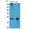
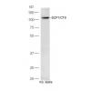
Fig2: Western blot analysis of CD1C against HEK293 (1) and CD1C (AA: extra 18-302)-hIgGFc transfected HEK293 (2) cell lysate. Proteins were transferred to a PVDF membrane and blocked with 5% BSA in PBS for 1 hour at room temperature. The primary antibody was used in 5% BSA at room temperature for 2 hours. Goat Anti-Mouse IgG - HRP Secondary Antibody at 1:5,000 dilution was used for 1 hour at room temperature.
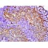
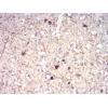
Fig3: Immunohistochemical analysis of paraffin-embedded rectum cancer tissues using anti-CD1C antibody. The section was pre-treated using heat mediated antigen retrieval with Tris-EDTA buffer (pH 8.0) for 20 minutes. The tissues were blocked in 5% BSA for 30 minutes at room temperature, washed with ddH2O and PBS, and then probed with the primary antibody for 30 minutes at room temperature. The detection was performed using an HRP conjugated compact polymer system. DAB was used as the chromogen. Tissues were counterstained with hematoxylin and mounted with DPX.
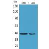
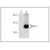
Fig4: Flow cytometric analysis of CD1C was done on Ramos cells. The cells were fixed, permeabilized and stained with the primary antibody(green). After incubation of the primary antibody at room temperature for an hour, the cells were stained with a Alexa Fluor 488-conjugated goat anti-Mouse IgG Secondary antibody at 1/500 dilution for 30 minutes. Unlabelled sample was used as a control (cells without incubation with primary antibody; red).
特别提示:本公司的所有产品仅可用于科研实验,严禁用于临床医疗及其他非科研用途!

![Anti-CD1C antibody [3G1B3]](images/no_picture.gif)