Anti-TNFRSF18 antibody [4H2D6]
-
概述
- 产品描述This gene encodes a member of the TNF-receptor superfamily. The encoded receptor has been shown to have increased expression upon T-cell activation, and it is thought to play a key role in dominant immunological self-tolerance maintained by CD25(+)CD4(+) regulatory T cells. Knockout studies in mice also suggest the role of this receptor is in the regulation of CD3-driven T-cell activation and programmed cell death. Three alternatively spliced transcript variants of this gene encoding distinct isoforms have been reported.
- 产品名称Anti-TNFRSF18 antibody [4H2D6]
- 分子量26kDa
- 种属反应性Human
- 验证应用WB,IHC-P,FC
- 抗体类型小鼠单抗
- 免疫原Purified recombinant fragment of human TNFRSF18 (AA: extra 26-162) expressed in E. Coli.
- 偶联Non-conjugated
-
性能
- 形态Liquid
- 浓度1 mg/mL
- 存放说明Store at +4℃ after thawing. Aliquot store at -20℃. Avoid repeated freeze / thaw cycles.
- 存储缓冲液1*PBS with 0.05% sodium azide.
- 亚型IgG1
- 纯化方式Protein G purified.
- 亚细胞定位Cell membrane
- 其它名称
- Activation-inducible TNFR family receptor
- Glucocorticoid-induced TNFR-related protein
- CD_antigen: CD357AITR, GITR
-
应用
WB: 1:500-1:2,000
IHC-P: 1:50-1:200
FC: 1:100-1:200
-
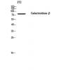
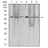
Fig1: Western blot analysis of TNFRSF18 against human TNFRSF18 (AA: extra 26-162) recombinant protein. Proteins were transferred to a PVDF membrane and blocked with 5% BSA in PBS for 1 hour at room temperature. The primary antibody (was used in 5% BSA at room temperature for 2 hours. Goat Anti-Mouse IgG - HRP Secondary Antibody at 1:5,000 dilution was used for 1 hour at room temperature.
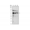
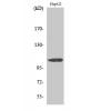
Fig2: Western blot analysis of against HEK293 (1) and TNFRSF18 (AA: extra 26-162)-hIgGFc transfected HEK293 (2) cell lysate.Proteins were transferred to a PVDF membrane and blocked with 5% BSA in PBS for 1 hour at room temperature. The primary antibody was used in 5% BSA at room temperature for 2 hours. Goat Anti-Mouse IgG - HRP Secondary Antibody at 1:5,000 dilution was used for 1 hour at room temperature.
Fig3: Immunohistochemical analysis of paraffin-embedded human cervical cancer tissue using anti-TNFRSF18 antibody. The section was pre-treated using heat mediated antigen retrieval with Tris-EDTA buffer (pH 8.0) for 20 minutes. The tissues were blocked in 5% BSA for 30 minutes at room temperature, washed with ddH2O and PBS, and then probed with the primary antibody for 30 minutes at room temperature. The detection was performed using an HRP conjugated compact polymer system. DAB was used as the chromogen. Tissues were counterstained with hematoxylin and mounted with DPX.
Fig4: Immunohistochemical analysis of paraffin-embedded human colon cancer tissue using anti-TNFRSF18 antibody. The section was pre-treated using heat mediated antigen retrieval with Tris-EDTA buffer (pH 8.0) for 20 minutes. The tissues were blocked in 5% BSA for 30 minutes at room temperature, washed with ddH2O and PBS, and then probed with the primary antibodyfor 30 minutes at room temperature. The detection was performed using an HRP conjugated compact polymer system. DAB was used as the chromogen. Tissues were counterstained with hematoxylin and mounted with DPX.
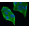
Fig5: Flow cytometric analysis of TNFRSF18 was done on Jurkat cells. The cells were fixed, permeabilized and stained with the primary antibody (green). After incubation of the primary antibody at room temperature for an hour, the cells were stained with a Alexa Fluor 488-conjugated goat anti-Mouse IgG Secondary antibody at 1/500 dilution for 30 minutes. Unlabelled sample was used as a control (cells without incubation with primary antibody; red).
特别提示:本公司的所有产品仅可用于科研实验,严禁用于临床医疗及其他非科研用途!

![Anti-TNFRSF18 antibody [4H2D6]](images/202012/goods_img/92571_G_1607495399426.jpg)