Anti-Myeloperoxidase antibody [A1F3]
-
概述
- 产品描述The heme protein myeloperoxidase (MPO) is a major component of azurophilic granules of neutrophils and polymorphonuclear leukocytes. Optimal oxygen-dependent microbiocidal activity depends on MPO as the critical enzyme for the generation of hypochlorous acid and other toxic oxygen products. The MPO precursor is synthesized during the promyelocytic stage of myeloid differentiation and is subsequently processed and transported intracellularly to the lysosomes. The precursor undergoes cotranslational N-linked glycosylation to produce a glycoprotein. Glucosidases in the endoplasmic reticulum (ER) or early cis Golgi convert the pro-MPO to a form which is sorted into a prelysosomal compartment, which undergoes final proteolytic maturation to native MPO, a pair of heavy-light protomers. In normal neutrophils, MPO is expressed as a dimer. Calreticulin, a calcium-binding protein residing in the ER, interacts specifically with fully glycosylated apopro-MPO. iMPO mRNA is abundant in human promyelocytic HL-60 and mouse myeloid leukemia NFS-60 cells. MPO is expressed at high levels in circulating neutrophils and monocytes but is not detectable in microglia, brain-specific macrophages or normal brain tissue.
- 产品名称Anti-Myeloperoxidase antibody [A1F3]
- 分子量51 kDa
- 种属反应性Human
- 验证应用WB,IHC-P,FC
- 抗体类型小鼠单抗
- 免疫原Recombinant protein within human Myeloperoxidase aa 550-745.
- 偶联Non-conjugated
-
性能
- 形态Liquid
- 浓度2 mg/mL
- 存放说明Store at +4℃ after thawing. Aliquot store at -20℃. Avoid repeated freeze / thaw cycles.
- 存储缓冲液1*PBS (pH7.4), 0.2% BSA, 50% Glycerol. Preservative: 0.05% Sodium Azide.
- 亚型IgG2a
- 纯化方式Protein G purified.
- 亚细胞定位Lysosome.
- 其它名称
- 84 kDa myeloperoxidase antibody
- 89 kDa myeloperoxidase antibody
- EC 1.11.1.7 antibody
more
-
应用
WB: 1:500-1:1,000
IHC-P: 1:50-1:200
FC: 1:50-1:100
-
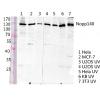
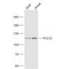
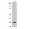
Fig1: Western blot analysis of Myeloperoxidase on HL-60 lysate. Proteins were transferred to a PVDF membrane and blocked with 5% BSA in PBS for 1 hour at room temperature. The primary antibody was used in 5% BSA at room temperature for 2 hours. Goat Anti-Mouse IgG - HRP Secondary Antibody (HA1006) at 1:5,000 dilution was used for 1 hour at room temperature.
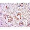
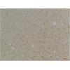
Fig2: Immunohistochemical analysis of paraffin-embedded human tonsil tissue using anti-Myeloperoxidase antibody. The section was pre-treated using heat mediated antigen retrieval with Tris-EDTA buffer (pH 8.0-8.4) for 20 minutes.The tissues were blocked in 5% BSA for 30 minutes at room temperature, washed with ddH2O and PBS, and then probed with the primary antibody for 30 minutes at room temperature. The detection was performed using an HRP conjugated compact polymer system. DAB was used as the chromogen. Tissues were counterstained with hematoxylin and mounted with DPX.
Fig3: Immunohistochemical analysis of paraffin-embedded human colon tissue using anti-Myeloperoxidase antibody. The section was pre-treated using heat mediated antigen retrieval with Tris-EDTA buffer (pH 8.0-8.4) for 20 minutes.The tissues were blocked in 5% BSA for 30 minutes at room temperature, washed with ddH2O and PBS, and then probed with the primary antibody for 30 minutes at room temperature. The detection was performed using an HRP conjugated compact polymer system. DAB was used as the chromogen. Tissues were counterstained with hematoxylin and mounted with DPX.
Fig4: Immunohistochemical analysis of paraffin-embedded human colon carcinoma tissue using anti-Myeloperoxidase antibody. The section was pre-treated using heat mediated antigen retrieval with Tris-EDTA buffer (pH 8.0-8.4) for 20 minutes.The tissues were blocked in 5% BSA for 30 minutes at room temperature, washed with ddH2O and PBS, and then probed with the primary antibody for 30 minutes at room temperature. The detection was performed using an HRP conjugated compact polymer system. DAB was used as the chromogen. Tissues were counterstained with hematoxylin and mounted with DPX.
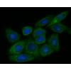
Fig5: Flow cytometric analysis of Myeloperoxidase was done on HL-60 cells. The cells were fixed, permeabilized and stained with the primary antibody (red). After incubation of the primary antibody at room temperature for an hour, the cells were stained with a Alexa Fluor 488-conjugated Goat anti-Mouse IgG Secondary antibody at 1/1000 dilution for 30 minutes.Unlabelled sample was used as a control (cells without incubation with primary antibody; black).
特别提示:本公司的所有产品仅可用于科研实验,严禁用于临床医疗及其他非科研用途!


![Anti-Myeloperoxidase antibody [A1F3]](images/202012/goods_img/92688_G_1607854064053.jpg)


