Anti-EHMT2 antibody [2D12A11]
-
概述
- 产品描述Histone methyltransferase that specifically mono- and dimethylates 'Lys-9' of histone H3 (H3K9me1 and H3K9me2, respectively) in euchromatin. H3K9me represents a specific tag for epigenetic transcriptional repression by recruiting HP1 proteins to methylated histones. Also mediates monomethylation of 'Lys-56' of histone H3 (H3K56me1) in G1 phase, leading to promote interaction between histone H3 and PCNA and regulating DNA replication. Also weakly methylates 'Lys-27' of histone H3 (H3K27me). Also required for DNA methylation, the histone methyltransferase activity is not required for DNA methylation, suggesting that these 2 activities function independently. Probably targeted to histone H3 by different DNA-binding proteins like E2F6, MGA, MAX and/or DP1. May also methylate histone H1. In addition to the histone methyltransferase activity, also methylates non-histone proteins: mediates dimethylation of 'Lys-373' of p53/TP53. Also methylates CDYL, WIZ, ACIN1, DNMT1, HDAC1, ERCC6, KLF12 and itself.
- 产品名称Anti-EHMT2 antibody [2D12A11]
- 分子量132.4kDa
- 种属反应性Human
- 验证应用WB,IHC-P,ICC,FC
- 抗体类型小鼠单抗
- 免疫原Purified recombinant fragment of human EHMT2 (AA: 317-471) expressed in E. Coli.
- 偶联Non-conjugated
-
性能
- 形态Liquid
- 浓度1 mg/mL
- 存放说明Store at +4℃ after thawing. Aliquot store at -20℃. Avoid repeated freeze / thaw cycles.
- 存储缓冲液1*PBS with 0.05% sodium azide.
- 亚型IgG1
- 纯化方式Protein G purified.
- 亚细胞定位Nucleus. Chromosome. Associates with euchromatic regions. Does not associate with heterochromatin.
- 其它名称
- Ankyrin repeat containing protein antibody
- Bat 8 antibody
- Bat8 antibody
more
-
应用
WB: 1:500-1:2,000
IHC-P: 1:50-1:200
ICC: 1:50-1:200
FC: 1:100-1:200
-
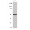
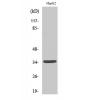
Fig1: Western blot analysis of EHMT2 against human EHMT2 (AA: 317-471) recombinant protein. Proteins were transferred to a PVDF membrane and blocked with 5% BSA in PBS for 1 hour at room temperature. The primary antibody was used in 5% BSA at room temperature for 2 hours. Goat Anti-Mouse IgG - HRP Secondary Antibody at 1:5,000 dilution was used for 1 hour at room temperature.
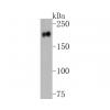
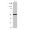
Fig2: Western blot analysis of EHMT2 against HEK293 (1) and EHMT2 (AA: 317-471)-hIgGFc transfected HEK293 (2) cell lysate. Proteins were transferred to a PVDF membrane and blocked with 5% BSA in PBS for 1 hour at room temperature. The primary antibody was used in 5% BSA at room temperature for 2 hours. Goat Anti-Mouse IgG - HRP Secondary Antibody at 1:5,000 dilution was used for 1 hour at room temperature.
Fig3: Immunocytochemistry staining of EHMT2 in Hela cells (green). Formalin fixed cells were permeabilized with 0.1% Triton X-100 in TBS for 10 minutes at room temperature and blocked with 1% Blocker BSA for 15 minutes at room temperature. Cells were probed with the primary antibody for 1 hour at room temperature, washed with PBS. Alexa Fluor®488 Goat anti-Mouse IgG was used as the secondary antibody at 1/1,000 dilution. Actin filaments have been labeled with Alexa Fluor- 555 phalloidin (red).
Fig4: Immunohistochemical analysis of paraffin-embedded bladder cancer tissues using anti-EHMT2 antibody. The section was pre-treated using heat mediated antigen retrieval with Tris-EDTA buffer (pH 8.0) for 20 minutes. The tissues were blocked in 5% BSA for 30 minutes at room temperature, washed with ddH2O and PBS, and then probed with the primary antibody ( for 30 minutes at room temperature. The detection was performed using an HRP conjugated compact polymer system. DAB was used as the chromogen. Tissues were counterstained with hematoxylin and mounted with DPX.
Fig5: Immunohistochemical analysis of paraffin-embedded rectum cancer tissues using anti-EHMT2 antibody. The section was pre-treated using heat mediated antigen retrieval with Tris-EDTA buffer (pH 8.0) for 20 minutes. The tissues were blocked in 5% BSA for 30 minutes at room temperature, washed with ddH2O and PBS, and then probed with the primary antibody for 30 minutes at room temperature. The detection was performed using an HRP conjugated compact polymer system. DAB was used as the chromogen. Tissues were counterstained with hematoxylin and mounted with DPX.
Fig6: Flow cytometric analysis of EHMT2 was done on HL-60 cells. The cells were fixed, permeabilized and stained with the primary antibody (green). After incubation of the primary antibody at room temperature for an hour, the cells were stained with a Alexa Fluor 488-conjugated goat anti-Mouse IgG Secondary antibody at 1/500 dilution for 30 minutes. Unlabelled sample was used as a control (cells without incubation with primary antibody; red).
特别提示:本公司的所有产品仅可用于科研实验,严禁用于临床医疗及其他非科研用途!

![Anti-EHMT2 antibody [2D12A11]](images/202012/goods_img/92721_G_1607858189208.jpg)