Anti-PER3 antibody [9B-7D]
-
概述
- 产品描述Biological timepieces called circadian clocks are responsible for the regulation of hormonal rhythms, sleep cycles and other behaviors. The superchiasmatic nucleus (SCN), which is located in the brain, was the first mammalian circadian clock to be discovered. A number of transcription factors appearing to be molecular components of the SCN clock have been identified. Mutations within the Clock gene increase the length of the endogenous period and cause a loss of rhythmicity of circadian oscillations. Three mammalian period proteins, designated Per1, Per2 and Per3, exhibit circadian rhythyms in the SCN. During subjective night, Per1 and Per2 RNA levels increase in response to light pulses while Per3 RNA levels show no change in response to light pulses. Tim, for timeless, interacts with Per1 as well as Per2; and Tim and Per1 negatively regulate Clock-BMAL1-induced transcription.
- 产品名称Anti-PER3 antibody [9B-7D]
- 分子量132 kDa
- 种属反应性Human
- 验证应用WB,IHC-P,ICC,FC
- 抗体类型小鼠单抗
- 免疫原Recombinant protein
- 偶联Non-conjugated
-
性能
- 形态Liquid
- 浓度2 mg/mL.
- 存放说明Store at +4℃ after thawing. Aliquot store at -20℃ or -80℃. Avoid repeated freeze / thaw cycles.
- 存储缓冲液1*TBS (pH7.4), 1%BSA, 40%Glycerol. Preservative: 0.05% Sodium Azide.
- 亚型IgG2b
- 纯化方式Protein A purified.
- 亚细胞定位Cytoplasm, nucleus
- 其它名称
- 2810049O06Rik antibody
- Cell growth inhibiting gene 13 protein antibody
- Cell growth-inhibiting gene 13 protein antibody
more
-
应用
WB: 1:500-1:2,000
ICC: 1:50-1:200
IHC-P: 1:50-1:200
FC: 1:50-1:100
-
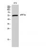
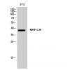
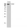
Fig1: Western blot analysis of PER3 on human PER3 recombinant protein using anti-PER3 antibody at 1/1,000 dilution.
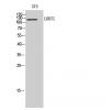
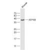
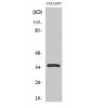
Fig2: Western blot analysis of PER3 on HEK293 (1) and PER3-hIgGFc transfected HEK293 (2) cell lysate using anti-PER3 antibody at 1/1,000 dilution.
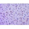
Fig3: ICC staining PER3 (green) and Actin filaments (red) in Hela cells. The nuclear counter stain is DAPI (blue). Cells were fixed in paraformaldehyde, permeabilised with 0.25% Triton X100/PBS.
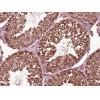
Fig4: Immunohistochemical analysis of paraffin-embedded human ovarian cancer tissue using anti-PER3 antibody. Counter stained with hematoxylin.
Fig5: Immunohistochemical analysis of paraffin-embedded human rectum cancer tissue using anti-PER3 antibody. Counter stained with hematoxylin.
Fig6: Flow cytometric analysis of Hela cells with PER3 antibody at 1/100 dilution (green) compared with an unlabelled control (cells without incubation with primary antibody; red).
特别提示:本公司的所有产品仅可用于科研实验,严禁用于临床医疗及其他非科研用途!

![Anti-PER3 antibody [9B-7D]](images/202012/goods_img/92753_G_1607942002250.jpg)