Anti-CLEC4F antibody [10-A6-1]
-
概述
- 产品描述CLEC4F, a member of C-type lectin, was first purified from rat liver extract with high binding affinity to fucose, galactose (Gal), N-acetylgalactosamine (GalNAc), and un-sialylated glucosphingolipids with GalNAc or Gal terminus. CLEC4F is a heavily glycosylated membrane protein co-expressed with F4/80 on Kupffer cells. In contrast to F4/80, CLEC4F is detectable in fetal livers at embryonic day 11.5 (E11.5) but not in yolk sac, suggesting the expression of CLEC4F is induced as cells migrate from yolk cells to the liver. Even though CLEC4F is not detectable in tissues outside liver, both residential Kupffer cells and infiltrating mononuclear cells surrounding liver abscesses are CLEC4F-positive upon Listeria monocytogenes (L. monocytogenes) infection. While CLEC4F has strong binding to Gal and GalNAc, terminal fucosylation inhibits CLEC4F recognition to several glycans such as Fucosyl GM1, Globo H, Bb3∼4 and other fucosyl-glycans. Moreover, CLEC4F interacts with alpha-galactosylceramide (α-GalCer) in a calcium-dependent manner and participates in the presentation of α-GalCer to natural killer T (NKT) cells. This suggests that CLEC4F is a C-type lectin with diverse binding specificity expressed on residential Kupffer cells and infiltrating monocytes in the liver, and may play an important role to modulate glycolipids presentation on Kupffer cells.
- 产品名称Anti-CLEC4F antibody [10-A6-1]
- 分子量Predicted band size 65 kDa.
- 种属反应性Human
- 验证应用WB,FC
- 抗体类型小鼠单抗
- 免疫原Recombinant protein within human CLEC4F aa 90-500.
- 偶联Non-conjugated
-
性能
- 形态Liquid
- 浓度2 mg/mL.
- 存放说明Store at +4℃ after thawing. Aliquot store at -20℃. Avoid repeated freeze / thaw cycles.
- 存储缓冲液1*PBS (pH7.4), 0.2% BSA, 50% Glycerol. Preservative: 0.05% Sodium Azide.
- 亚型IgG1
- 纯化方式Protein A affinity purified.
- 亚细胞定位Membrane.
- 其它名称
- C type (calcium dependent, carbohydrate recognition domain) lectin, superfamily member 13 antibody
- C-type lectin 13 antibody
- C-type lectin domain family 4 member F antibody
more
-
应用
WB:1:500-1:2,000
FC:1:50-1:100
-
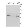
Fig1: Western blot analysis of CLEC4F on SH-SY5Y cell lysates. Proteins were transferred to a PVDF membrane and blocked with 5% BSA in PBS for 1 hour at room temperature. The primary antibody) was used in 5% BSA at room temperature for 2 hours. Goat Anti-Mouse IgG - HRP Secondary Antibody (HA1006) at 1:5,000 dilution was used for 1 hour at room temperature.
Fig2: Flow cytometric analysis of CLEC4F was done on SH-SY5Y cells. The cells were fixed, permeabilized and stained with the primary antibody (red). After incubation of the primary antibody at room temperature for an hour, the cells were stained with a Alexa Fluor 488-conjugated Goat anti-Mouse IgG Secondary antibody at 1/1000 dilution for 30 minutes.Unlabelled sample was used as a control (cells without incubation with primary antibody; black).
特别提示:本公司的所有产品仅可用于科研实验,严禁用于临床医疗及其他非科研用途!

![Anti-CLEC4F antibody [10-A6-1]](images/202012/goods_img/92935_G_1608203626894.jpg)