Anti-Cytokeratin 19 antibody [A3D1]
-
概述
- 产品描述The keratin gene family is a large group of intermediate filament and structural proteins that provide the structural components of hair, nails, horns, scales, claws and similar types of hard tissues. Keratins are also important for intracellular stability in epithelial tissues and different keratins often display organ- or tissue specific expression patterns. Due to these specific expression patterns, keratins are often used as diagnostic biomarkers. Keratin 19 is a type I keratin and expressed in simple epithelia including glandular cell types present in the GI-tract, female tissues, male tissues, and respiratory epithelium. Clinically, keratin 19 is used together with keratin 18 to distinguish hepatocellular cancer from cholangiocellular carcinoma (both keratins are expressed in bile ducts, but keratin 18 is expressed in hepatocytes whereas keratin 19 is not). Keratin 19 is a type I keratin. The type I cytokeratins consist of acidic proteins which are arranged in pairs of heterotypic keratin chains. Unlike its related family members, this smallest known acidic cytokeratin is not paired with a basic cytokeratin in epithelial cells. It is specifically found in the periderm, the transiently superficial layer that envelops the developing epidermis. The type I cytokeratins are clustered in a region of chromosome 17q12-q21.
- 产品名称Anti-Cytokeratin 19 antibody [A3D1]
- 分子量44 kDa
- 种属反应性Human
- 验证应用WB,IHC-P,FC
- 抗体类型小鼠单抗
- 免疫原Recombinant protein within Human Keratin 19 aa 128-322.
- 偶联Non-conjugated
-
性能
- 形态Liquid
- 浓度1 mg/ml.
- 存放说明Store at +4℃ after thawing. Aliquot store at -20℃. Avoid repeated freeze / thaw cycles.
- 存储缓冲液1*PBS (pH7.4), 0.2% BSA, 50% Glycerol. Preservative: 0.05% Sodium Azide.
- 亚型IgG1
- 纯化方式Protein A purified.
- 亚细胞定位Cytoskeleton, Cytosol.
- 其它名称
- 40 kDa keratin intermediate filament antibody
- CK 19 antibody
- CK-19 antibody
more
-
应用
WB: 1:2000-1:10000
IHC-P: 1:500-1:2000
FC: 1:50-1:100
-
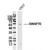
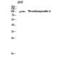
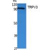
Fig1: Western blot analysis of Cytokeratin 19 on MCF-7 cell lysate. Proteins were transferred to a PVDF membrane and blocked with 5% BSA in PBS for 1 hour at room temperature. The primary antibodywas used in 5% BSA at room temperature for 2 hours. Goat Anti-Rabbit IgG - HRP Secondary Antibody (HA1001) at 1:5,000 dilution was used for 1 hour at room temperature.
Fig2: Immunohistochemical analysis of paraffin-embedded Human breast cancer tissue using anti-Cytokeratin 19 antibody. The section was pre-treated using heat mediated antigen retrieval with Tris-EDTA buffer (pH 8.0-8.4) for 20 minutes.The tissues were blocked in 5% BSA for 30 minutes at room temperature, washed with ddH2O and PBS, and then probed with the primary antibody for 30 minutes at room temperature. The detection was performed using an HRP conjugated compact polymer system. DAB was used as the chromogen. Tissues were counterstained with hematoxylin and mounted with DPX.
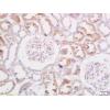
Fig3: Immunohistochemical analysis of paraffin-embedded Human small intestine tissue using anti-Cytokeratin 19 antibody. The section was pre-treated using heat mediated antigen retrieval with Tris-EDTA buffer (pH 8.0-8.4) for 20 minutes.The tissues were blocked in 5% BSA for 30 minutes at room temperature, washed with ddH2O and PBS, and then probed with the primary antibody for 30 minutes at room temperature. The detection was performed using an HRP conjugated compact polymer system. DAB was used as the chromogen. Tissues were counterstained with hematoxylin and mounted with DPX.
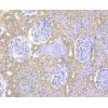
Fig4: Immunohistochemical analysis of paraffin-embedded Human pancreas tissue using anti-Cytokeratin 19 antibody. The section was pre-treated using heat mediated antigen retrieval with Tris-EDTA buffer (pH 8.0-8.4) for 20 minutes.The tissues were blocked in 5% BSA for 30 minutes at room temperature, washed with ddH2O and PBS, and then probed with the primary antibody for 30 minutes at room temperature. The detection was performed using an HRP conjugated compact polymer system. DAB was used as the chromogen. Tissues were counterstained with hematoxylin and mounted with DPX.
Fig5: Immunohistochemical analysis of paraffin-embedded Human Colon cancer tissue using anti-Cytokeratin 19 antibody. The section was pre-treated using heat mediated antigen retrieval with Tris-EDTA buffer (pH 8.0-8.4) for 20 minutes.The tissues were blocked in 5% BSA for 30 minutes at room temperature, washed with ddH2O and PBS, and then probed with the primary antibody for 30 minutes at room temperature. The detection was performed using an HRP conjugated compact polymer system. DAB was used as the chromogen. Tissues were counterstained with hematoxylin and mounted with DPX.
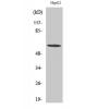
Fig6: Flow cytometric analysis of Cytokeratin 19 was done on MCF-7 cells. The cells were fixed, permeabilized and stained with the primary antibody (red). After incubation of the primary antibody at room temperature for an hour, the cells were stained with a Alexa Fluor 488-conjugated goat anti-rabbit IgG Secondary antibody at 1/500 dilution for 30 minutes.Unlabelled sample was used as a control (cells without incubation with primary antibody; black).
特别提示:本公司的所有产品仅可用于科研实验,严禁用于临床医疗及其他非科研用途!


![Anti-Cytokeratin 19 antibody [A3D1]](images/202012/goods_img/92945_G_1608204408372.jpg)


