Anti-CCL3 antibody
-
概述
- 产品描述Chemokines are members of a superfamily of small inducible, secreted, pro-inflammatory cytokines. Members of the chemokine family exhibit 20 to 50% homology in their predicted amino acid sequences and are divided into four subfamilies. In C-C (or b) subfamily, the first two cysteines are adjacent. C-C chemokines are chemoattractants and activators for monocytes and T cells. C-C subfamily members include macrophage inflammatory protein (MIP)-1α, MIP-1β, MIP-2, MIP-3α, MIP-3β, MIP-4, HCC-1, MIP-5 (or HCC-2), RANTES, MCP-1/2/3 (and the murine homologs JE and MARC), I-309, murine C10 and TCA3. Research has shown that MIP-1β is more selective than MIP-1α, primarily attracting CD4+ T lymphocytes, with a preference for T cells of the naive phenotype. MIP-1α is a more potent lymphocyte chemoattractant than MIP-1β and exhibits a broader range of chemoattractant specificities. It has been suggested that CD8+ T lymphocytes are involved in the control of HIV infection in vivo by the release of HIV-suppressive factors (HIV-SF). MIP-1α has been identified as one of the major HIV-SFs produced by CD8+ T cells, along with MIP-1β and RANTES
- 产品名称Anti-CCL3 antibody
- 种属反应性Human,Mouse,Rat
- 验证应用ICC,IHC-P,FC,ELISA
- 抗体类型兔多抗
- 免疫原Synthetic peptide (KLH-coupled) within mouse CCL3 50-80 aa.
- 偶联Non-conjugated
-
性能
- 形态Liquid
- 浓度1 mg/mL.
- 存放说明Store at +4℃ after thawing. Aliquot store at -20℃ or -80℃. Avoid repeated freeze / thaw cycles.
- 存储缓冲液1*PBS (pH7.4), 0.2% BSA, 40% Glycerol. Preservative: 0.05% Sodium Azide.
- 亚型IgG
- 纯化方式Peptide affinity purified
- 亚细胞定位Secreted
- 其它名称
- C C motif chemokine 3 antibody
- CCL 3 antibody
- CCL3 antibody
more
-
应用
ICC: 1:50-1:200
IHC-P: 1:50-1:200
FC:1:50-1:100
-
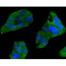
Fig1: Immunocytochemical staining of Hela cells using anti-CCL3 rabbit polyclonal antibody.
Fig2: Immunocytochemical staining of PANC-1 cells using anti-CCL3 rabbit polyclonal antibody.
Fig3: Immunocytochemical staining of A549 cells using anti-CCL3 rabbit polyclonal antibody.
Fig4: Immunohistochemical analysis of paraffin- embedded mouse lung tissue using anti-CCL3 rabbit polyclonal antibody.
Fig5: Immunohistochemical analysis of paraffin- embedded mouse spleen tissue using anti-CCL3 rabbit polyclonal antibody.
Fig6: Immunohistochemical analysis of paraffin- embedded mouse pancreas tissue using anti-CCL3 rabbit polyclonal antibody.
Fig7: Flow cytometric analysis of Hela cells with CCL3 antibody at 1/50 dilution (red) compared with an unlabelled control (cells without incubation with primary antibody; black). Alexa Fluor 488-conjugated Goat anti rabbit IgG was used as the secondary a
特别提示:本公司的所有产品仅可用于科研实验,严禁用于临床医疗及其他非科研用途!