Anti-IL17A antibody
-
概述
- 产品描述Interleukin 17A (IL-17 or IL-17A) is a pro-inflammatory cytokine. This cytokine is produced by a group of T helper cell known as T helper 17 cell in response to their stimulation with IL-23. Originally, Th17 was identified in 1993 by Rouvier et al. who isolated IL17 transcript from a rodent T-cell hybridoma. The protein encoded by IL17A is a founding member of IL-17 family (see below). IL17 protein exhibits a high homology with a viral IL-17-like protein encoded in the genome of T-lymphotropic rhadinovirus Herpesvirus saimiri.[2] In rodents, IL-17 is often referred to as CTLA8.The biologically active IL-17 interacts with type I cell surface receptor IL-17R. In turn, there are at least three variants of IL-17R referred to as IL17RA, IL17RB, and IL17RC. After binding to the receptor, IL-17 activates several signalling cascades that, in turn, lead to the induction of chemokines. Acting as chemoattractants, these chemokines recruit the immune cells, such as monocytes and neutrophils to the site of inflammation. Typically, the signaling events mentioned above follow an invasion of the body by pathogens. Promoting the inflammation, IL-17 acts in concert with tumor necrosis factor and interleukin-1.Moreover, an activation of IL-17 signalling is often observed in the pathogenesis of various autoimmune disorders, such as psoriasis.
- 产品名称Anti-IL17A antibody
- 分子量Predicted band size 17 kDa.
- 种属反应性Human,Mouse,Rat
- 验证应用WB,IHC-P,FC
- 抗体类型兔多抗
- 免疫原Recombinant protein within human IL17 aa 1-155.
- 偶联Non-conjugated
-
性能
- 形态Liquid
- 浓度1 mg/ml.
- 存放说明Store at +4℃ after thawing. Aliquot store at -20℃. Avoid repeated freeze / thaw cycles.
- 存储缓冲液1*PBS (pH7.4), 0.2% BSA, 50% Glycerol. Preservative: 0.05% Sodium Azide.
- 亚型IgG
- 纯化方式Protein affinity purified.
- 亚细胞定位Secreted.
- 其它名称
- CTLA 8 antibody
- CTLA-8 antibody
- CTLA8 antibody
more
-
应用
WB: 1:500
IHC-P: 1:50-1:100
FC: 1:50-1:100
-
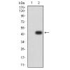
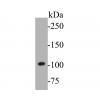
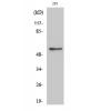
Fig1: Western blot analysis of IL17A on recombinant protein lysate. Proteins were transferred to a PVDF membrane and blocked with 5% BSA in PBS for 1 hour at room temperature. The primary antibody was used in 5% BSA at room temperature for 2 hours. Goat Anti-Rabbit IgG - HRP Secondary Antibody (HA1001) at 1:5,000 dilution was used for 1 hour at room temperature.
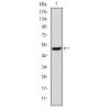
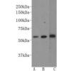
Fig2: Western blot analysis of IL17A on Rat stomach tissue lysate. Proteins were transferred to a PVDF membrane and blocked with 5% BSA in PBS for 1 hour at room temperature. The primary antibody was used in 5% BSA at room temperature for 2 hours. Goat Anti-Rabbit IgG - HRP Secondary Antibody (HA1001) at 1:5,000 dilution was used for 1 hour at room temperature.
Fig3: Western blot analysis of IL17A on Human skeletal muscle tissue lysate. Proteins were transferred to a PVDF membrane and blocked with 5% BSA in PBS for 1 hour at room temperature. The primary antibody was used in 5% BSA at room temperature for 2 hours. Goat Anti-Rabbit IgG - HRP Secondary Antibody (HA1001) at 1:5,000 dilution was used for 1 hour at room temperature.
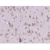
Fig4: Immunohistochemical analysis of paraffin-embedded Human skeletal muscle tissue using anti-IL17A antibody. The section was pre-treated using heat mediated antigen retrieval with Tris-EDTA buffer (pH 8.0-8.4) for 20 minutes.The tissues were blocked in 5% BSA for 30 minutes at room temperature, washed with ddH2O and PBS, and then probed with the primary antibodyfor 30 minutes at room temperature. The detection was performed using an HRP conjugated compact polymer system. DAB was used as the chromogen. Tissues were counterstained with hematoxylin and mounted with DPX.
Fig5: Immunohistochemical analysis of paraffin-embedded Human kidney tissue using anti-IL17A antibody. The section was pre-treated using heat mediated antigen retrieval with Tris-EDTA buffer (pH 8.0-8.4) for 20 minutes.The tissues were blocked in 5% BSA for 30 minutes at room temperature, washed with ddH2O and PBS, and then probed with the primary antibody for 30 minutes at room temperature. The detection was performed using an HRP conjugated compact polymer system. DAB was used as the chromogen. Tissues were counterstained with hematoxylin and mounted with DPX.
Fig6: Immunohistochemical analysis of paraffin-embedded mouse colon tissue using anti-IL17A antibody. The section was pre-treated using heat mediated antigen retrieval with Tris-EDTA buffer (pH 8.0-8.4) for 20 minutes.The tissues were blocked in 5% BSA for 30 minutes at room temperature, washed with ddH2O and PBS, and then probed with the primary antibody for 30 minutes at room temperature. The detection was performed using an HRP conjugated compact polymer system. DAB was used as the chromogen. Tissues were counterstained with hematoxylin and mounted with DPX.
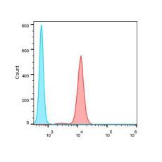
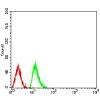
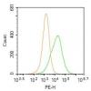
Fig7: Flow cytometric analysis of IL17A was done on Jurkat cells. The cells were fixed, permeabilized and stained with the primary antibody (red). After incubation of the primary antibody at room temperature for an hour, the cells were stained with a Alexa Fluor 488-conjugated goat anti-rabbit IgG Secondary antibody at 1/500 dilution for 30 minutes.Unlabelled sample was used as a control (cells without incubation with primary antibody; blue).
特别提示:本公司的所有产品仅可用于科研实验,严禁用于临床医疗及其他非科研用途!