Anti-Histone H4 antibody [H2-E7]
-
概述
- 产品描述In eukaryotes, DNA is wrapped around histone octamers to form the basic unit of chromatin structure. The octamer is composed of histones H2A, H2B, H3 and H4, and it associates with approximately 200 base pairs of DNA to form the nucleosome. The association of DNA with histones results in dense packing of chromatin, which restricts proteins involved in gene transcription from binding to DNA. p300 preferentially acetylates Histone H3 at lysines 14 and 18 and Histone H4 at lysines 5 and 8. PCAF in its native form, primarily acetylates Histone H3 at lysine 14 to a monoacetylated form, and less efficiently acetylates Histone H4 at lysine 8. Histone H4 may also be acetylated at lysines 12 and 16, and the involvement of acetylated H4 with Histones H2A, H2B and H3 suggests that acetylated histones may be involved in dynamic chromatin remodeling.
- 产品名称Anti-Histone H4 antibody [H2-E7]
- 分子量11.4 kDa
- 种属反应性Human
- 验证应用WB,ICC,IHC-P,FC
- 抗体类型小鼠单抗
- 免疫原Recombinant protein
- 偶联Non-conjugated
-
性能
- 形态Liquid
- 浓度2 mg/mL.
- 存放说明Store at +4℃ after thawing. Aliquot store at -20℃ or -80℃. Avoid repeated freeze / thaw cycles.
- 存储缓冲液1*TBS (pH7.4), 1%BSA, 40%Glycerol. Preservative: 0.05% Sodium Azide.
- 亚型IgG1
- 纯化方式Protein A purified.
- 亚细胞定位Nucleus. Chromosome.
- 其它名称
- dJ160A22.1 antibody
- dJ160A22.2 antibody
- dJ221C16.1 antibody
more
-
应用
WB: 1:500-1:2,000
ICC: 1:200-1:500
IHC-P: 1:200-1:500
FC: 1:100-1:200
-
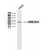
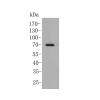
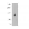
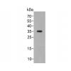
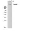
Fig1: Western blot analysis of Histone H4 on THP-1 cell lysate using anti- Histone H4 antibody at 1/1,000 dilution.
Fig2: ICC staining Histone H4 (green) and Actin filaments (red) in HeLa cells. The nuclear counter stain is DAPI (blue). Cells were fixed in paraformaldehyde, permeabilised with 0.25% Triton X100/PBS.
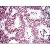
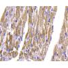
Fig3: Immunohistochemical analysis of paraffin-embedded human stomach cancer tissues using anti- Histone H4 antibody.
Fig4: Immunohistochemical analysis of paraffin-embedded human rectum cancer tissues using anti- Histone H4 antibody.
Fig5: Flow cytometric analysis of Raji cells with Histone H4 antiboy at 1/100 dilution (green) compared with an unlabelled control (cells without incubation with primary antibody; red).
特别提示:本公司的所有产品仅可用于科研实验,严禁用于临床医疗及其他非科研用途!


![Anti-Histone H4 antibody [H2-E7]](images/202012/goods_img/92558_G_1607494161026.jpg)


