Anti-Fascin antibody
-
概述
- 产品描述Actin-binding protein that contains 2 major actin binding sites. Organizes filamentous actin into parallel bundles. Plays a role in the organization of actin filament bundles and the formation of microspikes, membrane ruffles, and stress fibers. Important for the formation of a diverse set of cell protrusions, such as filopodia, and for cell motility and migration. Mediates reorganization of the actin cytoskeleton and axon growth cone collapse in response to NGF. Cell adhesion to extracellular matrix is an important physiological stimulus for organization of the Actin-based cytoskeleton. Adhesion to the matrix glycoprotein Thrombospondin 1 triggers the sustained formation of F-Actin microspikes that contain the Actin-bundling protein Fascin. These structures are also implicated in cell migration, which may be an important function of Thrombospondin 1 in tissue remodelling and wound repair. Fascin bundles Actin microfilaments within dynamic cellular structures such as microspikes, stress fibers and membrane ruffles. Fascin could serve as a prognostic factor for abnormal ovarian epithelial pathology and could be a novel target for the treatment of ovarian cancer. Fascin, an Actin-bundling protein, identifies dendritic cells in the blood and in tissues.
- 产品名称Anti-Fascin antibody
- 分子量54 kDa
- 种属反应性Human, Mouse, Rat
- 验证应用WB, IHC-P,FC
- 抗体类型兔多抗
- 免疫原Recombinant protein within Human Fascin aa 1-120.
- 偶联Non-conjugated
-
性能
- 形态Liquid
- 浓度1 mg/mL.
- 存放说明Store at +4℃ after thawing. Aliquot store at -20℃. Avoid repeated freeze / thaw cycles.
- 存储缓冲液1*PBS (pH7.4), 0.2% BSA, 50% Glycerol. Preservative: 0.05% Sodium Azide.
- 亚型IgG
- 纯化方式Protein affinity purified.
- 亚细胞定位Cytoskeleton. Cytosol.
- 其它名称
- 55 kDa actin bundling protein antibody
- 55 kDa actin-bundling protein antibody
- Actin bundling protein antibody
more
-
应用
WB:1:500-1:1,000
IHC-P:1:50-1:200
FC:1:50-1:100
-
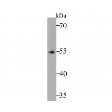
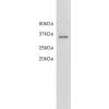
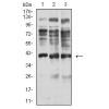
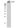
Fig1: Western blot analysis of Fascin on SH-SY5Y cell lysate. Proteins were transferred to a PVDF membrane and blocked with 5% BSA in PBS for 1 hour at room temperature. The primary antibody was used at a 1:500 dilution in 5% BSA at room temperature for 2 hours. Goat Anti-Rabbit IgG - HRP Secondary Antibody (HA1001) at 1:5,000 dilution was used for 1 hour at room temperature.
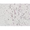
Fig2: Immunohistochemical analysis of paraffin-embedded rat brain tissue using anti-Fascin antibody. The section was pre-treated using heat mediated antigen retrieval with Tris-EDTA buffer (pH 8.0-8.4) for 20 minutes.The tissues were blocked in 5% BSA for 30 minutes at room temperature, washed with ddH2O and PBS, and then probed with the antibody at 1/50 dilution, for 30 minutes at room temperature and detected using an HRP conjugated compact polymer system. DAB was used as the chromogen. Counter stained with hematoxylin and mounted with DPX.
Fig3: Immunohistochemical analysis of paraffin-embedded human tonsil tissue using anti-Fascin antibody. The section was pre-treated using heat mediated antigen retrieval with Tris-EDTA buffer (pH 8.0-8.4) for 20 minutes.The tissues were blocked in 5% BSA for 30 minutes at room temperature, washed with ddH2O and PBS, and then probed with the antibody at 1/200 dilution, for 30 minutes at room temperature and detected using an HRP conjugated compact polymer system. DAB was used as the chromogen. Counter stained with hematoxylin and mounted with DPX.
Fig4: Immunohistochemical analysis of paraffin-embedded human kidney tissue using anti-Fascin antibody. The section was pre-treated using heat mediated antigen retrieval with Tris-EDTA buffer (pH 8.0-8.4) for 20 minutes.The tissues were blocked in 5% BSA for 30 minutes at room temperature, washed with ddH2O and PBS, and then probed with the antibody at 1/200 dilution, for 30 minutes at room temperature and detected using an HRP conjugated compact polymer system. DAB was used as the chromogen. Counter stained with hematoxylin and mounted with DPX.
Fig5: Immunohistochemical analysis of paraffin-embedded mouse testis tissue using anti-Fascin antibody. The section was pre-treated using heat mediated antigen retrieval with Tris-EDTA buffer (pH 8.0-8.4) for 20 minutes.The tissues were blocked in 5% BSA for 30 minutes at room temperature, washed with ddH2O and PBS, and then probed with the antibody at 1/200 dilution, for 30 minutes at room temperature and detected using an HRP conjugated compact polymer system. DAB was used as the chromogen. Counter stained with hematoxylin and mounted with DPX.
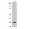
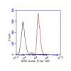
Fig6: Flow cytometric analysis of Fascin was done on SHSY5Y cells. The cells were fixed, permeabilized and stained with the primary antibody (, 1/50) (red). After incubation of the primary antibody at room temperature for an hour, the cells were stained with a Alexa Fluor 488-conjugated Goat anti-Rabbit IgG Secondary antibody at 1/1000 dilution for 30 minutes.Unlabelled sample was used as a control (cells without incubation with primary antibody; black).
特别提示:本公司的所有产品仅可用于科研实验,严禁用于临床医疗及其他非科研用途!