Anti-PTBP1 antibody [9A1]
-
概述
- 产品描述Plays a role in pre-mRNA splicing and in the regulation of alternative splicing events. Activates exon skipping of its own pre-mRNA during muscle cell differentiation. Binds to the polypyrimidine tract of introns. May promote RNA looping when bound to two separate polypyrimidine tracts in the same pre-mRNA. May promote the binding of U2 snRNP to pre-mRNA. Cooperates with RAVER1 to modulate switching between mutually exclusive exons during maturation of the TPM1 pre-mRNA. Represses the splicing of MAPT/Tau exon 10. In case of infection by picornaviruses, binds to the viral internal ribosome entry site (IRES) and stimulates the IRES-mediated translation.
- 产品名称Anti-PTBP1 antibody [9A1]
- 分子量57 kDa
- 种属反应性Human,Rat
- 验证应用WB,IHC-P,ICC,FC
- 抗体类型小鼠单抗
- 免疫原Recombinant protein within human PTBP1 aa 320-550.
- 偶联Non-conjugated
-
性能
- 形态Liquid
- 浓度2 mg/mL.
- 存放说明Store at +4℃ after thawing. Aliquot store at -20℃. Avoid repeated freeze / thaw cycles.
- 存储缓冲液1*PBS (pH7.4), 0.2% BSA, 50% Glycerol. Preservative: 0.05% Sodium Azide.
- 亚型IgG2a
- 纯化方式Protein G purified.
- 亚细胞定位Nucleus.
- 其它名称
- 57 kDa RNA binding protein PPTB 1 antibody
- 57 kDa RNA-binding protein PPTB-1 antibody
- Heterogeneous nuclear ribonucleoprotein I antibody
more
-
应用
WB: 1:5,000-1:10,000
IHC-P: 1:50-1:100
ICC: 1:50-1:100
FC: 1:50-1:100
-
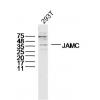
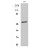
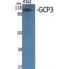
Fig1: Western blot analysis of PTBP1 on different cell lysates using anti-PTBP1 antibody at 1/5,000 dilution.
Positive control:
Lane 1: Jurkat
Lane 2: A431
Lane 3: K562
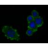
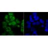
Fig2: ICC staining PTBP1 in A549 cells (green). The nuclear counter stain is DAPI (blue). Cells were fixed in paraformaldehyde, permeabilised with 0.25% Triton X100/PBS.
Fig3: ICC staining PTBP1 in 293T cells (green). The nuclear counter stain is DAPI (blue). Cells were fixed in paraformaldehyde, permeabilised with 0.25% Triton X100/PBS.
Fig4: ICC staining PTBP1 in SW480 cells (green). The nuclear counter stain is DAPI (blue). Cells were fixed in paraformaldehyde, permeabilised with 0.25% Triton X100/PBS.
Fig5: Immunohistochemical analysis of paraffin-embedded human kidney tissue using anti-PTBP1 antibody. Counter stained with hematoxylin.
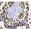
Fig6: Immunohistochemical analysis of paraffin-embedded human tonsil tissue using anti-PTBP1 antibody. Counter stained with hematoxylin.
Fig7: Immunohistochemical analysis of paraffin-embedded rat testis tissue using anti-PTBP1 antibody. Counter stained with hematoxylin.
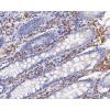
Fig8: Immunohistochemical analysis of paraffin-embedded human colon cancer tissue using anti-PTBP1 antibody. Counter stained with hematoxylin.
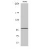
Fig9: Flow cytometric analysis of K562 cells with PTBP1 antibody at 1/100 dilution (red) compared with an unlabelled control (cells without incubation with primary antibody; black). Alexa Fluor 488-conjugated goat anti-mouse IgG was used as the secondary
特别提示:本公司的所有产品仅可用于科研实验,严禁用于临床医疗及其他非科研用途!

![Anti-PTBP1 antibody [9A1]](images/202012/goods_img/91997_G_1606895991874.jpg)