Anti-LAMP2 antibody [D3-D8-D3]
-
概述
- 产品描述Plays an important role in chaperone-mediated autophagy, a process that mediates lysosomal degradation of proteins in response to various stresses and as part of the normal turnover of proteins with a long biological half-live. Functions by binding target proteins, such as GAPDH and MLLT11, and targeting them for lysosomal degradation. Plays a role in lysosomal protein degradation in response to starvation (By similarity). Required for the fusion of autophagosomes with lysosomes during autophagy. Cells that lack LAMP2 express normal levels of VAMP8, but fail to accumulate STX17 on autophagosomes, which is the most likely explanation for the lack of fusion between autophagosomes and lysosomes. Required for normal degradation of the contents of autophagosomes. Required for efficient MHCII-mediated presentation of exogenous antigens via its function in lysosomal protein degradation; antigenic peptides generated by proteases in the endosomal/lysosomal compartment are captured by nascent MHCII subunits. Is not required for efficient MHCII-mediated presentation of endogenous antigens.
- 产品名称Anti-LAMP2 antibody [D3-D8-D3]
- 分子量Predicted band size 45 kDa
- 种属反应性Human
- 验证应用WB,ICC,IHC-P,FC
- 抗体类型小鼠单抗
- 免疫原Recombinant protein within human LAMP2 aa 1-300.
- 偶联Non-conjugated
-
性能
- 形态Liquid
- 浓度2 mg/mL.
- 存放说明Store at +4℃ after thawing. Aliquot store at -20℃. Avoid repeated freeze / thaw cycles.
- 存储缓冲液1*PBS (pH7.4), 0.2% BSA, 50% Glycerol. Preservative: 0.05% Sodium Azide.
- 亚型IgG1
- 纯化方式Protein G affinity purified.
- 亚细胞定位Cell membrane, lysosome membrane, endosome membrane, autophagosome membrane.
- 其它名称
- CD107 antigen like family member B antibody
- CD107 antigen-like family member B antibody
- CD107b antibody
more
-
应用
WB: 1:500-1:2,000
ICC: 1:50-1:100
IHC-P: 1:50-1:200
FC:1: 50-1:100
-
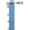
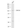
Fig1: Western blot analysis of LAMP2 on SiHa cell lysates. Proteins were transferred to a PVDF membrane and blocked with 5% BSA in PBS for 1 hour at room temperature. The primary antibody was used in 5% BSA at room temperature for 2 hours. Goat Anti-Mouse IgG - HRP Secondary Antibody (HA1006) at 1:5,000 dilution was used for 1 hour at room temperature.
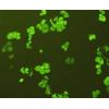
Fig2: ICC staining of LAMP2 in A549 cells (green). Formalin fixed cells were permeabilized with 0.1% Triton X-100 in TBS for 10 minutes at room temperature and blocked with 1% Blocker BSA for 15 minutes at room temperature. Cells were probed with the primary antibody for 1 hour at room temperature, washed with PBS. Alexa Fluor®488 Goat anti-Mouse IgG was used as the secondary antibody at 1/1,000 dilution. The nuclear counter stain is DAPI (blue).
Fig3: ICC staining of LAMP2 in HT-29 cells (green). Formalin fixed cells were permeabilized with 0.1% Triton X-100 in TBS for 10 minutes at room temperature and blocked with 1% Blocker BSA for 15 minutes at room temperature. Cells were probed with the primary antibody for 1 hour at room temperature, washed with PBS. Alexa Fluor®488 Goat anti-Mouse IgG was used as the secondary antibody at 1/1,000 dilution. The nuclear counter stain is DAPI (blue).
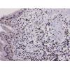
Fig4: Immunohistochemical analysis of paraffin-embedded human liver carcinoma tissue using anti-LAMP2 antibody. The section was pre-treated using heat mediated antigen retrieval with Tris-EDTA buffer (pH 8.0-8.4) for 20 minutes.The tissues were blocked in 5% BSA for 30 minutes at room temperature, washed with ddH2O and PBS, and then probed with the primary antibody for 30 minutes at room temperature. The detection was performed using an HRP conjugated compact polymer system. DAB was used as the chromogen. Tissues were counterstained with hematoxylin and mounted with DPX.
Fig5: Immunohistochemical analysis of paraffin-embedded human prostate tissue using anti-LAMP2 antibody. The section was pre-treated using heat mediated antigen retrieval with Tris-EDTA buffer (pH 8.0-8.4) for 20 minutes.The tissues were blocked in 5% BSA for 30 minutes at room temperature, washed with ddH2O and PBS, and then probed with the primary antibody for 30 minutes at room temperature. The detection was performed using an HRP conjugated compact polymer system. DAB was used as the chromogen. Tissues were counterstained with hematoxylin and mounted with DPX.
Fig6: Immunohistochemical analysis of paraffin-embedded human placenta tissue using anti-LAMP2 antibody. The section was pre-treated using heat mediated antigen retrieval with Tris-EDTA buffer (pH 8.0-8.4) for 20 minutes.The tissues were blocked in 5% BSA for 30 minutes at room temperature, washed with ddH2O and PBS, and then probed with the primary antibody for 30 minutes at room temperature. The detection was performed using an HRP conjugated compact polymer system. DAB was used as the chromogen. Tissues were counterstained with hematoxylin and mounted with DPX.
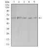
Fig7: Flow cytometric analysis of LAMP2 was done on SiHa cells. The cells were fixed, permeabilized and stained with the primary antibody (red). After incubation of the primary antibody at room temperature for an hour, the cells were sta
特别提示:本公司的所有产品仅可用于科研实验,严禁用于临床医疗及其他非科研用途!

![Anti-LAMP2 antibody [D3-D8-D3]](images/202012/goods_img/92377_G_1607252145013.jpg)