Anti-APEX1 antibody [G7-A2]
-
概述
- 产品描述The role of transcription factors in the regulation of gene expression is well established. Although the activity of these factors can be regulated by phosphorylation, evidence has indicated regulation of DNA binding mediated by changes in reduction-oxidation (redox) status. Mutational analysis has identified a single conserved cysteine residue mapping within the DNA binding domains of Fos and Jun. Chemical oxidation or modification of this cysteine residue inhibits the DNA binding activity of Fos and Jun. A similar mode of regulation has been recently proposed for other nuclear transcription factors. Oxidation is reversible by these compounds or by a cellular redox/DNA repair protein identified originally as Ref-1 (redox factor 1). Ref-1 is identical to a previously characterized DNA repair enzyme designated HAP1, APE or APEX.
- 产品名称Anti-APEX1 antibody [G7-A2]
- 分子量31 kDa
- 种属反应性Human,Monkey,Rat
- 验证应用WB,IHC-P,FC
- 抗体类型小鼠单抗
- 免疫原Recombinant protein
- 偶联Non-conjugated
-
性能
- 形态Liquid
- 浓度2 mg/mL.
- 存放说明Store at +4℃ after thawing. Aliquot store at -20℃ or -80℃. Avoid repeated freeze / thaw cycles.
- 存储缓冲液1*TBS (pH7.4), 1%BSA, 40%Glycerol. Preservative: 0.05% Sodium Azide.
- 亚型IgG1
- 纯化方式Protein A purified.
- 亚细胞定位Nucleus. Mitochondrion.?
- 其它名称
- AP endonuclease 1 antibody
- AP endonuclease class I antibody
- AP lyase antibody
more
-
应用
WB: 1:500-1:2,000
IHC-P: 1:100-1:500
FC: 1:100-1:200
-
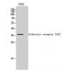
Fig1: Western blot analysis of APEX1 on human APEX1 recombinant protein using anti-APEX1 antibody at 1/1,000 dilution.
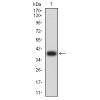
Fig2: Western blot analysis of APEX1 on HEK293 (1) and APEX1-hIgGFc transfected HEK293 (2) cell lysate using anti-APEX1 antibody at 1/1,000 dilution.
Fig3: Western blot analysis of APEX1 on different cell lysate using anti-APEX1 antibody at 1/1,000 dilution.
Positive control:
Lane 1: Hela
Lane 2: Jurkat
Lane 3: SW480
Lane 4: A431
Lane 5: HepG2
Lane 6: NIH/3T3
Lane 7: PC-12
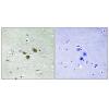
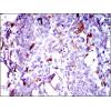
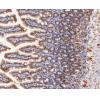
Fig4: Immunohistochemical analysis of paraffin-embedded human breast cancer tissue using anti-APEX1 antibody. Counter stained with hematoxylin.
Fig5: Immunohistochemical analysis of paraffin-embedded human esophageal cancer tissue using anti-APEX1 antibody. Counter stained with hematoxylin.
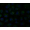
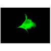
Fig6: Flow cytometric analysis of Hela cells with APEX1 antibody at 1/100 dilution (green) compared with an unlabelled control (cells without incubation with primary antibody; red).
Fig7: Flow cytometric analysis of SK-N-SH cells with APEX1 antibody at 1/100 dilution (green) compared with an unlabelled control (cells without incubation with primary antibody; red).
特别提示:本公司的所有产品仅可用于科研实验,严禁用于临床医疗及其他非科研用途!

![Anti-APEX1 antibody [G7-A2]](images/202012/goods_img/92136_G_1606985772766.jpg)