Anti-MSH6 antibody [A4C4]
-
概述
- 产品描述This gene encodes a member of the DNA mismatch repair MutS family. In E. coli, the MutS protein helps in the recognition of mismatched nucleotides prior to their repair. A highly conserved region of approximately 150 aa, called the Walker-A adenine nucleotide binding motif, exists in MutS homologs. The encoded protein heterodimerizes with MSH2 to form a mismatch recognition complex that functions as a bidirectional molecular switch that exchanges ADP and ATP as DNA mismatches are bound and dissociated. Mutations in this gene may be associated with hereditary nonpolyposis colon cancer, colorectal cancer, and endometrial cancer. Transcripts variants encoding different isoforms have been described.
- 产品名称Anti-MSH6 antibody [A4C4]
- 分子量153 kDa
- 种属反应性Human,Mouse,Rat
- 验证应用WB,IHC-P,FC,IF
- 抗体类型小鼠单抗
- 免疫原Synthetic peptide corresponding to C-terminal Human MSH6.
- 偶联Non-conjugated
-
性能
- 形态Liquid
- 浓度2 mg/mL.
- 存放说明Store at +4℃ after thawing. Aliquot store at -20℃. Avoid repeated freeze / thaw cycles.
- 存储缓冲液1*PBS (pH7.4), 0.2% BSA, 50% Glycerol. Preservative: 0.05% Sodium Azide.
- 亚型IgG2b
- 纯化方式Protein G affinity purified.
- 亚细胞定位Nucleus, Chromosome.
- 其它名称
- DNA mismatch repair protein Msh6 antibody
- G/T mismatch binding protein antibody
- G/T mismatch-binding protein antibody
more
-
应用
WB:1:500-1:2,000
IF: 1:50-1:100
IHC-P:1:50-1:200
FC:1:50-1:100
-
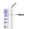
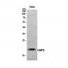
Fig1: Western blot analysis of MSH6 on different lysates. Proteins were transferred to a PVDF membrane and blocked with 5% BSA in PBS for 1 hour at room temperature. The primary antibody ( was used in 5% BSA at room temperature for 2 hours. Goat Anti-Mouse IgG - HRP Secondary Antibody (HA1006) at 1:5,000 dilution was used for 1 hour at room temperature.
Positive control:
Lane 1: human skin tissue lysate
Lane 2: rat brain tissue lysate
Fig2: Immunofluorescence staining of paraffin- embedded human breast carcinoma using anti-Rubisco activase rabbit polyclonal antibody.The section was pre-treated using heat mediated antigen retrieval with Tris-EDTA buffer (pH 9.0) for 20 minutes.(sodium citrate buffer (pH6) for 20 mins.) The tissues were blocked in 10% negative goat serum for 1 hour at room temperature, washed with PBS, and then probed with 1/50 dilution for 10 hours at 4℃ and detected using Alexa Fluor® 488 conjugate-Goat anti-Rabbit IgG (H+L) Secondary Antibody at a dilution of 1:500 for 1 hour at room temperature.
Fig3: Immunofluorescence staining of paraffin- embedded mouse testis using anti-Rubisco activase rabbit polyclonal antibody.The section was pre-treated using heat mediated antigen retrieval with Tris-EDTA buffer (pH 9.0) for 20 minutes.(sodium citrate buffer (pH6) for 20 mins.) The tissues were blocked in 10% negative goat serum for 1 hour at room temperature, washed with PBS, and then probed with at 1/50 dilution for 10 hours at 4℃ and detected using Alexa Fluor® 488 conjugate-Goat anti-Rabbit IgG (H+L) Secondary Antibody at a dilution of 1:500 for 1 hour at room temperature.
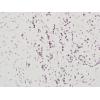
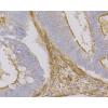
Fig4: Immunohistochemical analysis of paraffin-embedded human thyroid tissue using anti-MSH6 antibody. The section was pre-treated using heat mediated antigen retrieval with sodium citrate buffer (pH 6.0) for 20 minutes. The tissues were blocked in 5% BSA for 30 minutes at room temperature, washed with ddH2O and PBS, and then probed with the primary antibody ( for 30 minutes at room temperature. The detection was performed using an HRP conjugated compact polymer system. DAB was used as the chromogen. Tissues were counterstained with hematoxylin and mounted with DPX.
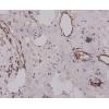
Fig5: Immunohistochemical analysis of paraffin-embedded human colon carcinoma tissue using anti-MSH6 antibody. The section was pre-treated using heat mediated antigen retrieval with sodium citrate buffer (pH 6.0) for 20 minutes. The tissues were blocked in 5% BSA for 30 minutes at room temperature, washed with ddH2O and PBS, and then probed with the primary antibody for 30 minutes at room temperature. The detection was performed using an HRP conjugated compact polymer system. DAB was used as the chromogen. Tissues were counterstained with hematoxylin and mounted with DPX.
Fig6: Immunohistochemical analysis of paraffin-embedded human skin tissue using anti-MSH6 antibody. The section was pre-treated using heat mediated antigen retrieval with sodium citrate buffer (pH 6.0) for 20 minutes. The tissues were blocked in 5% BSA for 30 minutes at room temperature, washed with ddH2O and PBS, and then probed with the primary antibody ( for 30 minutes at room temperature. The detection was performed using an HRP conjugated compact polymer system. DAB was used as the chromogen. Tissues were counterstained with hematoxylin and mounted with DPX.
Fig7: Immunohistochemical analysis of paraffin-embedded human breast carcinoma tissue using anti-MSH6 antibody. The section was pre-treated using heat mediated antigen retrieval with sodium citrate buffer (pH 6.0) for 20 minutes. The tissues were blocked in 5% BSA for 30 minutes at room temperature, washed with ddH2O and PBS, and then probed with the primary antibody for 30 minutes at room temperature. The detection was performed using an HRP conjugated compact polymer system. DAB was used as the chromogen. Tissues were counterstained with hematoxylin and mounted with DPX.
Fig8: Immunohistochemical analysis of paraffin-embedded human esophagus tissue using anti-MSH6 antibody. The section was pre-treated using heat mediated antigen retrieval with sodium citrate buffer (pH 6.0) for 20 minutes. The tissues were blocked in 5% BSA for 30 minutes at room temperature, washed with ddH2O and PBS, and then probed with the primary antibody for 30 minutes at room temperature. The detection was performed using an HRP conjugated compact polymer system. DAB was used as the chromogen. Tissues were counterstained with hematoxylin and mounted with DPX.
Fig9: Immunohistochemical analysis of paraffin-embedded human placenta tissue using anti-MSH6 antibody. The section was pre-treated using heat mediated antigen retrieval with sodium citrate buffer (pH 6.0) for 20 minutes. The tissues were blocked in 5% BSA for 30 minutes at room temperature, washed with ddH2O and PBS, and then probed with the primary antibody for 30 minutes at room temperature. The detection was performed using an HRP conjugated compact polymer system. DAB was used as the chromogen. Tissues were counterstained with hematoxylin and mounted with DPX.
Fig10: Immunohistochemical analysis of paraffin-embedded mouse testis tissue using anti-MSH6 antibody. The section was pre-treated using heat mediated antigen retrieval with sodium citrate buffer (pH 6.0) for 20 minutes. The tissues were blocked in 5% BSA for 30 minutes at room temperature, washed with ddH2O and PBS, and then probed with the primary antibody for 30 minutes at room temperature. The detection was performed using an HRP conjugated compact polymer system. DAB was used as the chromogen. Tissues were counterstained with hematoxylin and mounted with DPX.
Fig11: Immunohistochemical analysis of paraffin-embedded mouse colon tissue using anti-MSH6 antibody. The section was pre-treated using heat mediated antigen retrieval with sodium citrate buffer (pH 6.0) for 20 minutes. The tissues were blocked in 5% BSA for 30 minutes at room temperature, washed with ddH2O and PBS, and then probed with the primary antibody for 30 minutes at room temperature. The detection was performed using an HRP conjugated compact polymer system. DAB was used as the chromogen. Tissues were counterstained with hematoxylin and mounted with DPX.
Fig12: Immunohistochemical analysis of paraffin-embedded mouse placenta tissue using anti-MSH6 antibody. The section was pre-treated using heat mediated antigen retrieval with sodium citrate buffer (pH 6.0) for 20 minutes. The tissues were blocked in 5% BSA for 30 minutes at room temperature, washed with ddH2O and PBS, and then probed with the primary antibody (for 30 minutes at room temperature. The detection was performed using an HRP conjugated compact polymer system. DAB was used as the chromogen. Tissues were counterstained with hematoxylin and mounted with DPX.
Fig13: Flow cytometric analysis of MSH6 was done on K562 cells. The cells were fixed, permeabilized and stained with the primary antibody (red). After incubation of the primary antibody at room temperature for an hour, the cells were stained with a Alexa Fluor 488-conjugated Goat anti-Mouse IgG Secondary antibody at 1/1000 dilution for 30 minutes.Unlabelled sample was used as a control (cells without incubation with primary antibody; black).
特别提示:本公司的所有产品仅可用于科研实验,严禁用于临床医疗及其他非科研用途!

![Anti-MSH6 antibody [A4C4]](images/202012/goods_img/92258_G_1607170814134.jpg)