Anti-PTP1B antibody [D0-C7]
-
概述
- 产品描述The phosphorylation of proteins at tyrosine residues has long been recognized as an important regulatory component of signal transduction. This is a reversible process, involving both enzymes that phosphorylate proteins on tyrosine residues as well as a rapidly expanding family of protein tyrosine phosphatases. These latter enzymes bear little resemblance to either the protein serine and protein threonine phosphatases or to the acid and alkaline phosphatases. In most tissues, the major PTPase is a vanadate- and molybdate-sensitive protein. On the basis of sequence analysis, PTP1B (PTPase 1B) expressed in human placenta exhibits similarities both with the common leukocyte antigen (CD45) and with LAR, a homolog of the neural adhesion molecule (NCAM). PTP1B is synthesized as a 435 amino acid precursor protein which is cleaved to generate the active 321 amino acid enzyme.
- 产品名称Anti-PTP1B antibody [D0-C7]
- 分子量50kDa
- 种属反应性Human,Mouse,Rat
- 验证应用WB,ICC,FC
- 抗体类型小鼠单抗
- 免疫原recombinant protein
- 偶联Non-conjugated
-
性能
- 形态Liquid
- 浓度2 mg/mL.
- 存放说明Store at +4℃ after thawing. Aliquot store at -20℃ or -80℃. Avoid repeated freeze / thaw cycles.
- 存储缓冲液1*PBS (pH7.4), 0.2% BSA, 40% Glycerol. Preservative: 0.05% Sodium Azide.
- 亚型IgG1
- 纯化方式Protein A purified.
- 亚细胞定位Endoplasmic reticulum membrane
- 其它名称
- PTP1B antibody
- Non receptor tyrosine phosphatase 1 antibody
- Protein phosphotyrosylphosphatase 1B antibody
more
-
应用
WB: 1:1,000-1:2,000
ICC: 1:100-1:500
FC: 1:50-1:100
-
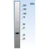
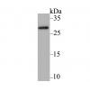
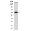
Fig1: Western blot analysis on different cell lysates using anti-PTP1B Mouse mAb. Positive control:
Lane 1: A431
Lane 2: Hela
Lane 3: HepG2
Lane 4: A549
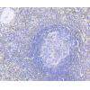
Fig2: Immunocytochemical staining of Hela cells using anti-PTP1B Mouse mAb.
Fig3: Immunocytochemical staining of A549 cells using anti-PTP1B Mouse mAb.
Fig4: Immunocytochemical staining of HepG2 cells using anti-PTP1B Mouse mAb.
Fig5: Flow cytometric analysis of Jurkat cells with PTP1B antibody at 1/100 dilution (red) compared with an unlabelled control (cells without incubation with primary antibody; black). Alexa Fluor 488-conjugated Goat anti mouse IgG was used as the secondary antibody
特别提示:本公司的所有产品仅可用于科研实验,严禁用于临床医疗及其他非科研用途!

![Anti-PTP1B antibody [D0-C7]](images/202012/goods_img/92346_G_1607249054603.jpg)