Anti-GRAMD1A antibody [1-E4]
-
概述
- 产品描述GRAMD1A first finds in human embryonic stem cell, and expresses in ectoderm, mesoderm and endoderm tissues and many tumor cells, its function hasn’t been explored. The researh has determined the role of GRAM1DA in the self-renewal of HCC stem cell and resistance to chemotherapy by overexpressing GRAMD1A, and the role of GRAMD1A in tumor growth by overexpressing or downregulating GRAMD1A. GRAMD1A is a prognostic factor for HCC, it promoted the self-renewal of HCC stem cell, resistance to chemotherapy and tumor growth through promoting STAT5, Inhibition of STAT5 in indicated cells with GRAMD1A overexpression inhibited the self-renewal of HCC stem cell, resistance to chemotherapy and tumor growth.
- 产品名称Anti-GRAMD1A antibody [1-E4]
- 分子量80 kDa
- 种属反应性Human,Mouse
- 验证应用WB,ICC,IHC-P
- 抗体类型小鼠单抗
- 免疫原Recombinant protein
- 偶联Non-conjugated
-
性能
- 形态Liquid
- 浓度2 mg/mL.
- 存放说明Store at +4℃ after thawing. Aliquot store at -20℃ or -80℃. Avoid repeated freeze / thaw cycles.
- 存储缓冲液1*PBS (pH7.4), 0.2% BSA, 40% Glycerol. Preservative: 0.05% Sodium Azide
- 亚型IgM
- 纯化方式Protein A purified.
- 亚细胞定位Membrane, nucleus.
-
应用
WB: 1:1,000-1:2,000
ICC: 1:50-1:200
IHC-P: 1:50-1:200
-
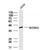
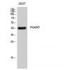
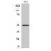
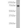
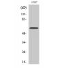
Fig1: Western blot analysis of GRAMD1A on NIH/3T3 (1) and SHG-44 (2) cells lysates using anti-GRAMD1A antibody at 1/1,000 dilution.
Fig2: ICC staining GRAMD1A in HepG2 cells (green). The nuclear counter stain is DAPI (blue). Cells were fixed in paraformaldehyde, permeabilised with 0.25% Triton X100/PBS.
Fig3: Immunohistochemical analysis of paraffin-embedded human tonsil tissue using anti-GRAMD1A antibody. Counter stained with hematoxylin.
Fig4: Immunohistochemical analysis of paraffin-embedded human endometrial cancer tissue using anti-GRAMD1A antibody. Counter stained with hematoxylin.
Fig5: Immunohistochemical analysis of paraffin-embedded human glioma tissue using anti-GRAMD1A antibody. Counter stained with hematoxylin.
Fig6: Immunohistochemical analysis of paraffin-embedded human colon tissue using anti-GRAMD1A antibody. Counter stained with hematoxylin.
Fig7: Immunohistochemical analysis of paraffin-embedded human renal cell carcinoma tissue using anti-GRAMD1A antibody. Counter stained with hematoxylin.
Fig8: Immunohistochemical analysis of paraffin-embedded human stamoch tissue using anti-GRAMD1A antibody. Counter stained with hematoxylin.
Fig9: Immunohistochemical analysis of paraffin-embedded human gastric adenocarcinoma tissue using anti-GRAMD1A antibody. Counter stained with hematoxylin.
特别提示:本公司的所有产品仅可用于科研实验,严禁用于临床医疗及其他非科研用途!

![Anti-GRAMD1A antibody [1-E4]](images/202012/goods_img/92420_G_1607328440441.jpg)