Anti-CSNK2B antibody [F3-F12]
-
概述
- 产品描述Casein kinase I (also designated CKI) and casein kinase II (CKII) compose a family of serine/threonine protein kinases which are present in all eukaryotes examined to date. Casein kinase I family members, which include casein kinase Iα, Iγ, Iδ and Ie, have been implicated in the control of cytoplasmic and nuclear processes, including DNA replication and repair. CKII is usually expressed as a tetrameric complex consisting of either an α2β2 or an αα'β2 structure. The a catalytic subunit is stimulated by the β regulatory subunit, which undergoes autophosphorylation. Casein kinase II activity is high in the cytosol and nucleus of proliferating and differentiating cells. Casein kinase II is known to phosphorylate more than 100 different substrates including nuclear oncoproteins, transcription factors and enzymes involved in DNA metabolism.
- 产品名称Anti-CSNK2B antibody [F3-F12]
- 分子量25 kDa
- 种属反应性Human
- 验证应用WB,ICC,IHC-P,FC
- 抗体类型小鼠单抗
- 免疫原Recombinant protein
- 偶联Non-conjugated
-
性能
- 形态Liquid
- 浓度2 mg/mL.
- 存放说明Store at +4℃ after thawing. Aliquot store at -20℃ or -80℃. Avoid repeated freeze / thaw cycles.
- 存储缓冲液1*TBS (pH7.4), 1%BSA, 40%Glycerol. Preservative: 0.05% Sodium Azide.
- 亚型IgG1
- 纯化方式Protein A purified.
- 亚细胞定位Chromatin, cytoplasm, cytosol, extracellular exosome, nuclear matrix, nucleoplasm,nucleus
- 其它名称
- Casein kinase 2 beta polypeptide antibody
- Casein kinase II beta subunit antibody
- Casein kinase II subunit beta antibody
more
-
应用
WB: 1:500-1:2,000
ICC: 1:50-1:200
IHC-P: 1:50-1:200
FC: 1:50-1:100
-
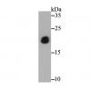
Fig1: Western blot analysis of CSNK2B on human CSNK2B recombinant protein using anti-CSNK2B antibody at 1/1,000 dilution.
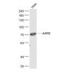
Fig2: Western blot analysis of CSNK2B on HEK293 (1) and CSNK2B-hIgGFc transfected HEK293 (2) cell lysate using anti-CSNK2B antibody at 1/1,000 dilution.
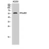
Fig3: Western blot analysis of CSNK2B on different cell lysates using anti-CSNK2B antibody at 1/1,000 dilution.
Positive control: Line1: Hela Line2: Jurkat Line3: K562 Line4: HepG2 Line5: C6 Line6: SK-N-SH Line7: NTERA-2 Line8: MCF-7 Line9: NIH/3T3
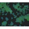
Fig4: ICC staining CSNK2B (green) and Actin filaments (red) in MCF-7 cells. The nuclear counter stain is DAPI (blue). Cells were fixed in paraformaldehyde, permeabilised with 0.25% Triton X100/PBS.
Fig5: Immunohistochemical analysis of paraffin-embedded human cervical cancer tissue using anti-CSNK2B antibody. Counter stained with hematoxylin.
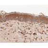
Fig6: Immunohistochemical analysis of paraffin-embedded human colon cancer tissue using anti-CSNK2B antibody. Counter stained with hematoxylin.
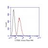
Fig7: Flow cytometric analysis of Hela cells with CSNK2B antibody at 1/100 dilution (green) compared with an unlabelled control (cells without incubation with primary antibody; red).
特别提示:本公司的所有产品仅可用于科研实验,严禁用于临床医疗及其他非科研用途!

![Anti-CSNK2B antibody [F3-F12]](images/202012/goods_img/92594_G_1607496958599.jpg)