Anti-EIF2A antibody [A8-A7]
-
概述
- 产品描述eIF2A (eukaryotic translation initiation factor 2A), also known as CDA02, MSTP004 or MSTP089, is a 585 amino acid protein that contains three WD repeats. Expressed ubiquitously with highest expression in heart, brain, pancreas and placenta, eIF2A functions as a translation initiation factor that binds Met-tRNA and directs it to 40S ribosomal subunits. Present in the early steps of protein synthesis, eIF2A controls the binding of Met-tRNA to 40S ribosomal subunits in a codon-dependent manner, in contrast to the eIF2 complex which accomplishes the same task in a GTP-dependent manner. In addition to its role in transcription initiation, eIF2A may also act to negatively regulate the expression of specific proteins, suggesting a possible role as a transcriptional repressor. eIF2A exists as two isoforms due to alternative splicing events.
- 产品名称Anti-EIF2A antibody [A8-A7]
- 分子量65 kDa
- 种属反应性Human
- 验证应用WB,ICC,IHC-P,FC
- 抗体类型小鼠单抗
- 免疫原Recombinant protein
- 偶联Non-conjugated
-
性能
- 形态Liquid
- 浓度2 mg/mL.
- 存放说明Store at +4℃ after thawing. Aliquot store at -20℃ or -80℃. Avoid repeated freeze / thaw cycles.
- 存储缓冲液1*TBS (pH7.4), 1%BSA, 40%Glycerol. Preservative: 0.05% Sodium Azide.
- 亚型IgG1
- 纯化方式Protein A purified.
- 亚细胞定位Cytoplasm.
- 其它名称
- 65 kDa eukaryotic translation initiation factor 2A antibody
- CDA 02 antibody
- CDA02 antibody
more
-
应用
WB: 1:500-1:2,000
ICC: 1:50-1:200
IHC-P: 1:50-1:200
FC: 1:50-1:100
-
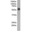
Fig1: Western blot analysis of EIF2A on human EIF2A recombinant protein using anti-EIF2A antibody at 1/1,000 dilution.
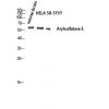
Fig2: Western blot analysis of EIF2A on HEK293 (1) and EIF2A-hIgGFc transfected HEK293 (2) cell lysate using anti-EIF2A antibody at 1/1,000 dilution.
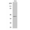
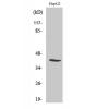
Fig3: Western blot analysis of EIF2A on different cell lysate using anti-EIF2A antibody at 1/1,000 dilution.
Positive control: Line1: MCF-7 Line2: PC-12 Line3: HepG2 Line4: Hela Line5: Cos7 Line6: K562 Line7: Jurkat Line8: A431 Line9:
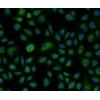
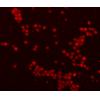
Fig4: ICC staining EIF2A (green) in HepG2 cells. The nuclear counter stain is DAPI (blue). Cells were fixed in paraformaldehyde, permeabilised with 0.25% Triton X100/PBS.
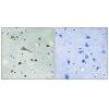
Fig5: Immunohistochemical analysis of paraffin-embedded human cervical cancer tissue using anti-EIF2A antibody. Counter stained with hematoxylin.
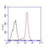
Fig6: Flow cytometric analysis of HepG2 cells with EIF2A antibody at 1/100 dilution (green) compared with an unlabelled control (cells without incubation with primary antibody; red).
特别提示:本公司的所有产品仅可用于科研实验,严禁用于临床医疗及其他非科研用途!

![Anti-EIF2A antibody [A8-A7]](images/202012/goods_img/92695_G_1607855084813.jpg)