Anti-IL3RA antibody [C5-E11]
-
概述
- 产品描述Interleukin-3, or IL-3, is a pleiotropic cytokine that is primarily secreted by activated T lymphocytes and stimulates the proliferation and differentiation of hematopoietic cells. IL-3 exerts its biological effects through a receptor which consists of a ligand-specific α subunit (IL-3Rα) and a signal transducing β subunit (IL-3Rβ) common to the IL-3/IL-5/GM-CSF receptors. The α subunits are low-affinity ligand-binding proteins while the β subunits do not themselves bind ligand, but are required for high affinity binding by the α subunits. The mouse IL-3 receptor has two distinct β subunits, one that functions only in IL-3-mediated cell signaling and a second that is shared with IL-5 and GM-CSF. The murine β subunits are 91% homologous at the amino acid level but only 56% homologous to the human β subunit. The carboxy-terminus of the β subunit has been shown to be necessary for activation of the MAP kinase signaling pathway. Although the IL-3 receptor has no intrinsic kinase activity, stimulation with IL-3 leads to tyrosine phosphorylation of the JAK/Tyk 2 family member, JAK2, which in turn activates and causes nuclear translocation of Stat5a and Stat5b.
- 产品名称Anti-IL3RA antibody [C5-E11]
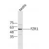
- 分子量43 kDa
- 种属反应性Human
- 验证应用WB,ICC,IHC-P,FC
- 抗体类型小鼠单抗
- 免疫原Recombinant protein
- 偶联Non-conjugated
-
性能
- 形态Liquid
- 浓度2 mg/mL.
- 存放说明Store at +4℃ after thawing. Aliquot store at -20℃ or -80℃. Avoid repeated freeze / thaw cycles.
- 存储缓冲液1*TBS (pH7.4), 1%BSA, Preservative: 0.05% Sodium Azide.
- 亚型IgG1
- 纯化方式Protein A purified.
- 亚细胞定位Membrane.
- 其它名称
- CD123 antibody
- CD123 antigen antibody
- hIL 3Ra antibody
more
-
应用
WB: 1:500-1:1,000
ICC: 1:50-1:200
IHC-P: 1:50-1:200
FC: 1:100-1:200
-
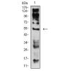
Fig1: Western blot analysis of IL3RA on human IL3RA recombinant protein using anti-IL3RA antibody at 1/1,000 dilution.
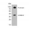
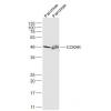
Fig2: Western blot analysis of IL3RA on HEK293 (1) and IL3RA-hIgGFc transfected HEK293 (2) cell lysate using anti-IL3RA antibody at 1/1,000 dilution.
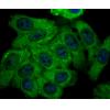
Fig3: ICC staining IL3RA (green) and Actin filaments (red) in Hela cells. The nuclear counter stain is DAPI (blue). Cells were fixed in paraformaldehyde, permeabilised with 0.25% Triton X100/PBS.
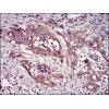
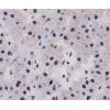
Fig4: Immunohistochemical analysis of paraffin-embedded human ovarian cancer tissue using anti-IL3RA antibody. Counter stained with hematoxylin.
Fig5: Immunohistochemical analysis of paraffin-embedded human lung cancer tissue using anti-IL3RA antibody. Counter stained with hematoxylin.
Fig6: Flow cytometric analysis of Hela cells with IL3RA antibody at 1/100 dilution (green) compared with an unlabelled control (cells without incubation with primary antibody; red).
特别提示:本公司的所有产品仅可用于科研实验,严禁用于临床医疗及其他非科研用途!

![Anti-IL3RA antibody [C5-E11]](images/202012/goods_img/92776_G_1608033057745.jpg)