Anti-Caveolin-1 antibody [A3E8]
-
概述
- 产品描述Caveolin-1 is a protein that in humans is encoded by the CAV1 gene. The scaffolding protein encoded by this gene is the main component of the caveolae plasma membranes found in most cell types. The protein links integrin subunits to the tyrosine kinase FYN, an initiating step in coupling integrins to the Ras-ERK pathway and promoting cell cycle progression. The gene is a tumor suppressor gene candidate and a negative regulator of the Ras-p42/44 MAP kinase cascade. CAV1 and CAV2 are located next to each other on chromosome 7 and express colocalizing proteins that form a stable hetero-oligomeric complex. By using alternative initiation codons in the same reading frame, two isoforms (alpha and beta) are encoded by a single transcript from this gene. Caveolin 1 has been shown to interact with heterotrimeric G proteins, Src tyrosine kinases (Src, Lyn) and H-Ras, cholesterol, TGF beta receptor 1, endothelial NOS, androgen receptor, amyloid precursor protein, gap junction protein, alpha 1, nitric oxide synthase 2A, epidermal growth factor receptor, endothelin receptor type B, PDGFRB, PDGFRA, PTGS2, TRAF2, estrogen receptor alpha, caveolin 2, PLD2, Bruton's tyrosine kinase and SCP2. All these interactions are through a caveolin-scaffolding domain (CSD) within caveolin-1 molecule. Molecules that interact with caveolin-1 contain caveolin-binding motifs (CBM).
- 产品名称Anti-Caveolin-1 antibody [A3E8]
- 分子量20 kDa
- 种属反应性Human
- 验证应用WB,ICC,IHC-P,FC
- 抗体类型小鼠单抗
- 免疫原Recombinant protein within human Caveolin-1 aa 1-150.
- 偶联Non-conjugated
-
性能
- 形态Liquid
- 浓度2 mg/mL.
- 存放说明Store at +4℃ after thawing. Aliquot store at -20℃. Avoid repeated freeze / thaw cycles.
- 存储缓冲液1*PBS (pH7.4), 0.2% BSA, 50% Glycerol. Preservative: 0.05% Sodium Azide.
- 亚型IgG2b
- 纯化方式Protein G affinity purified.
- 亚细胞定位Cell membrane, golgi apparatus membrane, trans-golgi network, caveola, membrane raft.
- 其它名称
- BSCL3 antibody
- CAV antibody
- CAV1 antibody
more
-
应用
WB:1:500-1:2,000
ICC:1:50-1:100
IHC-P:1:500-1:2,000
FC:1:50-1:100
-
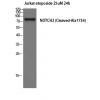
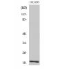
Fig1: Western blot analysis of Caveolin-1 on SiHa cell lysates. Proteins were transferred to a PVDF membrane and blocked with 5% BSA in PBS for 1 hour at room temperature. The primary antibody was used in 5% BSA at room temperature for 2 hours. Goat Anti-Mouse IgG - HRP Secondary Antibody (HA1006) at 1:5,000 dilution was used for 1 hour at room temperature.
Fig2: ICC staining of Caveolin-1 in SKOV-3 cells (green). Formalin fixed cells were permeabilized with 0.1% Triton X-100 in TBS for 10 minutes at room temperature and blocked with 1% Blocker BSA for 15 minutes at room temperature. Cells were probed with the primary antibody for 1 hour at room temperature, washed with PBS. Alexa Fluor®488 Goat anti-Mouse IgG was used as the secondary antibody at 1/1,000 dilution. The nuclear counter stain is DAPI (blue).
Fig3: Immunohistochemical analysis of paraffin-embedded human liver tissue using anti-Caveolin-1 antibody. The section was pre-treated using heat mediated antigen retrieval with Tris-EDTA buffer (pH 8.0-8.4) for 20 minutes.The tissues were blocked in 5% BSA for 30 minutes at room temperature, washed with ddH2O and PBS, and then probed with the primary antibody for 30 minutes at room temperature. The detection was performed using an HRP conjugated compact polymer system. DAB was used as the chromogen. Tissues were counterstained with hematoxylin and mounted with DPX.
Fig4: Immunohistochemical analysis of paraffin-embedded human lung tissue using anti-Caveolin-1 antibody. The section was pre-treated using heat mediated antigen retrieval with Tris-EDTA buffer (pH 8.0-8.4) for 20 minutes.The tissues were blocked in 5% BSA for 30 minutes at room temperature, washed with ddH2O and PBS, and then probed with the primary antibody ( for 30 minutes at room temperature. The detection was performed using an HRP conjugated compact polymer system. DAB was used as the chromogen. Tissues were counterstained with hematoxylin and mounted with DPX.
Fig5: Immunohistochemical analysis of paraffin-embedded human lung carcinoma tissue using anti-Caveolin-1 antibody. The section was pre-treated using heat mediated antigen retrieval with Tris-EDTA buffer (pH 8.0-8.4) for 20 minutes.The tissues were blocked in 5% BSA for 30 minutes at room temperature, washed with ddH2O and PBS, and then probed with the primary antibody for 30 minutes at room temperature. The detection was performed using an HRP conjugated compact polymer system. DAB was used as the chromogen. Tissues were counterstained with hematoxylin and mounted with DPX.
Fig6: Flow cytometric analysis of Caveolin-1 was done on PANC-1 cells. The cells were fixed, permeabilized and stained with the primary antibody ((red). After incubation of the primary antibody at room temperature for an hour, the cells were stained with a Alexa Fluor 488-conjugated Goat anti-Mouse IgG Secondary antibody at 1/1000 dilution for 30 minutes.Unlabelled sample was used as a control (cells without incubation with primary antibody; black)
特别提示:本公司的所有产品仅可用于科研实验,严禁用于临床医疗及其他非科研用途!


![Anti-Caveolin-1 antibody [A3E8]](images/202012/goods_img/92952_G_1608461491060.jpg)


