Anti-BubR1 antibody
-
概述
- 产品描述Human cells contain two related protein kinases, BUB1 and BUBR1, that appear to have evolved from a single ancestral BUB1 gene. Both kinases are concentrated near the surface of the kinetochore where they monitor kinetochore-microtubule interactions. BUB1 and BUBR1 bind to kinetochores and are postulated to be components of the mitotic checkpoint, which monitors kinetochore activities to determine if chromosomes have achieved alignment at the spindle equator. BUBR1 is essential for normal mitotic progression as it prevents cells from prematurely entering anaphase. BUB3 is a conserved component of the mitotic spindle assembly complex and is also involved with the essential spindle checkpoint pathway that operates during early embryogenesis.
- 产品名称Anti-BubR1 antibody
- 分子量119 kDa
- 种属反应性Human
- 验证应用WB,ICC,IHC-P,FC
- 抗体类型兔多抗
- 免疫原Recombinant protein within human BubR1 330-600 aa.
- 偶联Non-conjugated
-
性能
- 形态Liquid
- 浓度1 mg/mL.
- 存放说明Store at +4℃ after thawing. Aliquot store at -20℃. Avoid repeated freeze / thaw cycles.
- 存储缓冲液1*PBS (pH7.4), 0.2% BSA, 50% Glycerol. Preservative: 0.05% Sodium Azide.
- 亚型IgG
- 纯化方式Protein affinity purified.
- 亚细胞定位Nucleus. Cytoplasm.
- 其它名称Beta homolg of S. cerevisiae BUB 1 antibody
Beta homolg of S. cerevisiae budding uninhibited by benzimidazoles antibody
BUB 1B antibody
BUB1 budding uninhibited by benzimidazoles 1 homolog beta antibody
Bub1A antibody
BUB1B antibody
BUB1B_HUMAN antibody
BUB1beta antibody
BUBR1 antibody
Budding Uninhibited by Benzimidazoles 1 beta antibody
Budding uninhibited by benzimidazoles 1 homolog beta (yeast) antibody
hBUBR1 antibody
MAD3/BUB1 related protein kinase antibody
MAD3/BUB1-related protein kinase antibody
MAD3L antibody
Mitotic checkpoint gene BUB1B antibody
Mitotic checkpoint kinase MAD3L antibody
Mitotic checkpoint serine/threonine protein kinase BUB1 beta antibody
Mitotic checkpoint serine/threonine-protein kinase BUB1 beta antibody
MVA1 antibody
OTTHUMP00000160319 antibody
Protein SSK1 antibody
SSK 1 antibody
SSK1 antibody
more
-
应用
WB: 1:5,000-1:10,000
ICC: 1:50-1:200
IHC-P: 1:50-1:200
FC: 1:50-1:100
-
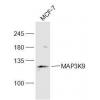
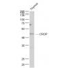
Fig1: Western blot analysis of BubR1 on SH-SY-5Y cell lysate using anti-BubR1 antibody at 1/10,000 dilution.
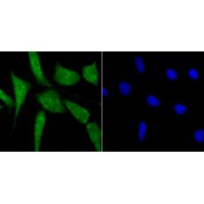
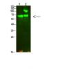
Fig2: ICC staining BubR1 in Hela cells (green). The nuclear counter stain is DAPI (blue). Cells were fixed in paraformaldehyde, permeabilised with 0.25% Triton X100/PBS.
Fig3: ICC staining BubR1 in SH-SY-5Y cells (green). The nuclear counter stain is DAPI (blue). Cells were fixed in paraformaldehyde, permeabilised with 0.25% Triton X100/PBS.
Fig4: ICC staining BubR1 in SiHa cells (green). The nuclear counter stain is DAPI (blue). Cells were fixed in paraformaldehyde, permeabilised with 0.25% Triton X100/PBS.
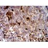
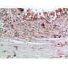
Fig5: Immunohistochemical analysis of paraffin-embedded human spleen tissue using anti-BubR1 antibody. Counter stained with hematoxylin.
Fig6: Flow cytometric analysis of HL-60 cells with BubR1 antibody at 1/100 dilution (red) compared with an unlabelled control (cells without incubation with primary antibody; black). Alexa Fluor 488-conjugated goat anti-rabbit IgG was used as the secondary antibody.
特别提示:本公司的所有产品仅可用于科研实验,严禁用于临床医疗及其他非科研用途!