Anti-LIM1 antibody
-
概述
- 产品描述The Lim gene family is a subfamily of homeobox genes. The homeobox genes are essential in organizing the body plan of an organism and all contain the same conserved homeodomain of amino acids.Evidence that Lim-1 is essential to a developing organism is its conservation throughout evolution and presence in a variety of organisms.The Lim-1 gene encodes a transcription factor which binds to the DNA of specific genes and functions to produce the needed gene product for development of the organism. Lim-1 is important during early molecular development and is required in both primitive streak-derived tissue and visceral endoderm of the early embryo for development of a head. Studies done using mutant organisms without the Lim gene results in organisms that develop no head structure at all support the essential role of the Lim-1 gene in formation of the head. This gene has also been shown to play a crucial role in the formation of the female reproductive tract.The gene is expressed in the developing Müllerian duct of females, and when the gene is knocked out no reproductive tract forms. Recent studies have shown that Lim-1 mutations may be one cause of the Mayer-Rokitansky-Küster-Hauser (MRKH) syndrome. MRKH is characterized by defective development, or absence, of the uterus and upper part of the vagina in women with normal ovaries and karyotype.
- 产品名称Anti-LIM1 antibody
- 分子量45 kDa
- 种属反应性Human,Mouse,Rat,Zebrafish
- 验证应用WB,IHC,FC
- 抗体类型兔多抗
- 免疫原Synthetic peptide within Zebrafish LIM1 aa 150-200.
- 偶联Non-conjugated
-
性能
- 形态Liquid
- 浓度1 mg/mL.
- 存放说明Store at +4℃ after thawing. Aliquot store at -20℃. Avoid repeated freeze / thaw cycles.
- 存储缓冲液1*PBS (pH7.4), 0.2% BSA, 50% Glycerol. Preservative: 0.05% Sodium Azide.
- 亚型IgG
- 纯化方式Peptide affinity purified.
- 亚细胞定位Nucleus.
- 其它名称
- hLim-1 antibody
- Homeo box protein Lim 1 antibody
- Homeo box protein Lim1 antibody
more
-
应用
WB: 1:500-1:1,000
IHC-P: 1:50-1:200
FC: 1:50-1:100
-
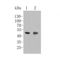
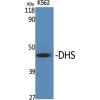
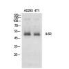
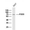
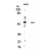
Fig1: Western blot analysis of LIM1 on different lysates. Proteins were transferred to a PVDF membrane and blocked with 5% BSA in PBS for 1 hour at room temperature. The primary antibody was used in 5% BSA at room temperature for 2 hours. Goat Anti-Rabbit IgG - HRP Secondary Antibody (HA1001) at 1:5,000 dilution was used for 1 hour at room temperature.
Positive control:
Lane 1: A431 cell lysate
Lane 2: Zebrafish tissue lysate
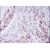
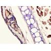
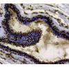
Fig2: Immunohistochemical analysis of paraffin-embedded rat brian tissue using anti-LIM1 antibody. The section was pre-treated using heat mediated antigen retrieval with sodium citrate buffer (pH 6.0) for 20 minutes. The tissues were blocked in 5% BSA for 30 minutes at room temperature, washed with ddH2O and PBS, and then probed with the primary antibody for 30 minutes at room temperature. The detection was performed using an HRP conjugated compact polymer system. DAB was used as the chromogen. Tissues were counterstained with hematoxylin and mounted with DPX.
Fig3: Immunohistochemical analysis of paraffin-embedded mouse brian tissue using anti-LIM1 antibody. The section was pre-treated using heat mediated antigen retrieval with sodium citrate buffer (pH 6.0) for 20 minutes. The tissues were blocked in 5% BSA for 30 minutes at room temperature, washed with ddH2O and PBS, and then probed with the primary antibody for 30 minutes at room temperature. The detection was performed using an HRP conjugated compact polymer system. DAB was used as the chromogen. Tissues were counterstained with hematoxylin and mounted with DPX.
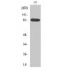
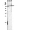
Fig4: Flow cytometric analysis of LIM1 was done on MCF-7 cells. The cells were fixed, permeabilized and stained with the primary antibody (red). After incubation of the primary antibody at room temperature for an hour, the cells were stained with a Alexa Fluor 488-conjugated goat anti-rabbit IgG Secondary antibody at 1/500 dilution for 30 minutes.Unlabelled sample was used as a control (cells without incubation with primary antibody; black).
特别提示:本公司的所有产品仅可用于科研实验,严禁用于临床医疗及其他非科研用途!