Anti-Hsc70 antibody
-
概述
- 产品描述The HSP 70 family is composed of four highly conserved proteins: HSP 70, HSC 70, GRP 75 and GRP 78. These proteins serve a variety of roles: they act as molecular chaperones facilitating the assembly of multi-protein complexes, participate in the translocation of polypeptides across cell membranes and to the nucleus, and aid in the proper folding of nascent polypeptide chains. All members of the family, except HSP 70, are constitutively expressed in primate cells. HSP 70 expression is strongly induced in response to heat stress. HSP 70 and HSC 70 play key roles in the cytosolic endoplasmic reticulum and mitochondrial import machinery and are found in both the cytosol and nucleus of mammalian cells. Both HSP 70 and HSC 70 are involved in the chaperoning of nascent polypeptide chains and in protecting cells against the accumulation of improperly folded proteins. GRP 78 is localized in the endoplasmic reticulum, where it receives imported secretory proteins and is involved in the folding and translocation of nascent peptide chains. GRP 75 expression is restricted to the mitochondrial matrix and aids in the translocation and folding of nascent polypeptide chains of both nuclear and mitochondrial origin. GRP 75 and GRP 78 are unresponsive to heat stress and are induced by glucose deprivation. It has been postulated that members of the HSP 70 family act as force-generating motors, relying on the hydrolysis of ATP for their activity.
- 产品名称Anti-Hsc70 antibody
- 分子量70 kDa
- 种属反应性Human,Mouse,Rat
- 验证应用WB,ICC,IHC-P,FC
- 抗体类型兔多抗
- 免疫原Recombinant protein.
- 偶联Non-conjugated
-
性能
- 形态Liquid
- 浓度1 mg/mL.
- 存放说明Store at +4℃ after thawing. Aliquot store at -20℃ or -80℃. Avoid repeated freeze / thaw cycles.
- 存储缓冲液1*PBS (pH7.4), 0.2% BSA, 50% Glycerol. Preservative: 0.05% Sodium Azide.
- 亚型IgG
- 纯化方式Protein A purified.
- 亚细胞定位Nucleus. Cytoplasm. Secreted.
- 其它名称
- 2410008N15Rik antibody
- Constitutive heat shock protein 70 antibody
- Epididymis luminal protein 33 antibody
more
-
应用
WB: 1:1,000-1:2,000
ICC: 1:50-1:200
IHC-P: 1:50-1:200
FC: 1:50-1:200
-
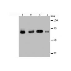
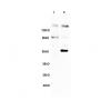
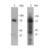
Fig1: Western blot analysis of Hsc70 on different cell lysate using anti-Hsc70 antibody at 1/2,000 dilution.
Positive control:
Lane 1: Hela Lane 2: A431 Lane 3: NIH-3T3 Lane 4: PC-12
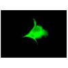
Fig2: ICC staining Hsc70 in Hela cells (red). The nuclear counter stain is DAPI (blue). Cells were fixed in paraformaldehyde, permeabilised with 0.25% Triton X100/PBS.
Fig3: ICC staining Hsc70 in A549 cells (green). The nuclear counter stain is DAPI (blue). Cells were fixed in paraformaldehyde, permeabilised with 0.25% Triton X100/PBS.
Fig4: ICC staining Hsc70 in SK-Br-3 cells (green). The nuclear counter stain is DAPI (blue). Cells were fixed in paraformaldehyde, permeabilised with 0.25% Triton X100/PBS.
Fig5: Immunohistochemical analysis of paraffin-embedded human tonsil tissue using anti-Hsc70 antibody. Counter stained with hematoxylin.
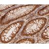
Fig6: Immunohistochemical analysis of paraffin-embedded human colon cancer tissue using anti-Hsc70 antibody. Counter stained with hematoxylin.
Fig7: Immunohistochemical analysis of paraffin-embedded human breast tissue using anti-Hsc70 antibody. Counter stained with hematoxylin.
Fig8: Immunohistochemical analysis of paraffin-embedded human kidney tissue using anti-Hsc70 antibody. Counter stained with hematoxylin.
Fig9: Immunohistochemical analysis of paraffin-embedded mouse testis tissue using anti-Hsc70 antibody. Counter stained with hematoxylin.
Fig10: Immunohistochemical analysis of paraffin-embedded mouse prostate tissue using anti-Hsc70 antibody. Counter stained with hematoxylin.
Fig11: Flow cytometric analysis of Jurkat cells with Hsc70 antibody at 1/100 dilution (red) compared with an unlabelled control (cells without incubation with primary antibody; black).
特别提示:本公司的所有产品仅可用于科研实验,严禁用于临床医疗及其他非科研用途!