Anti-Integrin beta 1 antibody [B10-A5]
-
概述
- 产品描述Integrins are transmembrane receptors that mediate the attachment between a cell and its surroundings, such as other cells or the extracellular matrix (ECM). Integrins are obligate heterodimers containing two distinct chains, called the α (alpha) and β (beta) subunits. The molecular mass of the integrin subunits can vary from 90 kDa to 160 kDa. Beta subunits have four cysteine-rich repeated sequences. Both α and β subunits bind several divalent cations. Integrins have two main functions: Attachment of the cell to the ECM and signal transduction from the ECM to the cell. However, they are also involved in a wide range of other biological activities, including immune patrolling, cell migration, and binding to cells by certain viruses, such as adenovirus, echovirus, hantavirus, and foot and mouth disease viruses. Research studies have implicated β1 integrin in various activities including embryonic development, blood vessel, skin, bone, and muscle formation, as well as tumor metastasis and angiogenesis.
- 产品名称Anti-Integrin beta 1 antibody [B10-A5]
- 分子量Predicted band size 88 kDa.
- 种属反应性Human, Mouse
- 验证应用WB,IHC-P,FC
- 抗体类型小鼠单抗
- 免疫原Synthetic peptide of the C-terminal Human Integrin beta 1.
- 偶联Non-conjugated
-
性能
- 形态Liquid
- 浓度2 mg/mL.
- 存放说明Store at +4℃ after thawing. Aliquot store at -20℃. Avoid repeated freeze / thaw cycles.
- 存储缓冲液1*PBS (pH7.4), 0.2% BSA, 50% Glycerol. Preservative: 0.05% Sodium Azide.
- 亚型IgM
- 纯化方式Protein A purified.
- 亚细胞定位Cell Membrane, Cell projection, Cleavage furrow.
- 其它名称
- beta1 integrin antibody
- CD29 antibody
- Fibronectin receptor subunit beta antibody
more
-
应用
WB: 1:1000-1:5000
IHC-P: 1:50-1:200
FC: 1:50-1:100
-
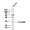
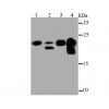
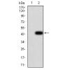
Fig1: Western blot analysis of Integrin beta 1 on different lysates. Proteins were transferred to a PVDF membrane and blocked with 5% BSA in PBS for 1 hour at room temperature. The primary antibody was used at a 1:1,000 dilution in 5% BSA at room temperature for 2 hours. Goat Anti-Mouse IgG - HRP Secondary Antibody (HA1001) at 1:5,000 dilution was used for 1 hour at room temperature.
Positive control:
Lane 1: Human liver tissue lysate, untreated
Lane 2: Mouse liver tissue lysate, untreated
Lane 2: A172 cell lysate, untreated
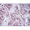
Fig2: Immunohistochemical analysis of paraffin-embedded human tonsil tissue using anti-Integrin beta 1 antibody. The section was pre-treated using heat mediated antigen retrieval with Tris-EDTA buffer (pH 8.0-8.4) for 20 minutes.The tissues were blocked in 5% BSA for 30 minutes at room temperature, washed with ddH2O and PBS, and then probed with the antibody at 1/200 dilution, for 30 minutes at room temperature and detected using an HRP conjugated compact polymer system. DAB was used as the chromogen. Counter stained with hematoxylin and mounted with DPX
Fig3: Immunohistochemical analysis of paraffin-embedded human kidney tissue using anti-Integrin beta 1 antibody. The section was pre-treated using heat mediated antigen retrieval with Tris-EDTA buffer (pH 8.0-8.4) for 20 minutes.The tissues were blocked in 5% BSA for 30 minutes at room temperature, washed with ddH2O and PBS, and then probed with the antibodyat 1/50 dilution, for 30 minutes at room temperature and detected using an HRP conjugated compact polymer system. DAB was used as the chromogen. Counter stained with hematoxylin and mounted with DPX
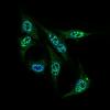
Fig4: Flow cytometric analysis of Integrin beta 1 was done on L929 cells. The cells were fixed, permeabilized and stained with the primary antibody (red). After incubation of the primary antibody at room temperature for an hour, the cells were stained with a Alexa Fluor 488-conjugated goat anti-rabbit IgG Secondary antibody at 1/500 dilution for 30 minutes.Unlabelled sample was used as a control (cells without incubation with primary antibody; black).
特别提示:本公司的所有产品仅可用于科研实验,严禁用于临床医疗及其他非科研用途!


![Anti-Integrin beta 1 antibody [B10-A5]](images/202012/goods_img/92465_G_1607331355041.jpg)


