Anti-DSG3 antibody [11G-2C]
-
概述
- 产品描述Pemphigus is an autoimmune disease of skin adhesion associated with auto-antibodies against a number of keratinocyte antigens, such as the adhesion molecules desmoglein (dsg) 1 and 3 and acetylcholine receptors. Desmogleins, type I membrane proteins, are important for cell adhesion and are expressed in great abundance at the desmosomes, which are adhesive cell junctions. Desmogleins belong to the cadherin family and consist of dsg1, dsg2 and dsg3. Calcium binds to the putative calcium binding sites at the extracellular N-terminal domain, which has cadherin-like repeats. Unlike normal human keratinocytes, the squamous cell carcinoma cells exhibit diminished or un-usual expression of dsg3 and dsg1, which bear pemphigus vulgaris and pemphigus foliaceus antigens, respectively. Several carcinoma cell lines constantly express dsg2 and dsg3 mRNA, whereas cultured normal human keratinocytes always express dsg1 and dsg3 mRNA, with or without dsg2 mRNA. This expression pattern indicates that desmoglein isoforms exhibit abnormal expression and may be related to tumor cell kinetics, such as cell invasion and metastasis. dsg2 is the fundamental dsg common to all desmosome-possessing tissues and is the largest desmoglein in the family.
- 产品名称Anti-DSG3 antibody [11G-2C]
- 分子量108 kDa
- 种属反应性Human
- 验证应用WB,IHC-P,FC
- 抗体类型小鼠单抗
- 免疫原Recombinant protein
- 偶联Non-conjugated
-
性能
- 形态Liquid
- 浓度2 mg/mL.
- 存放说明Store at +4℃ after thawing. Aliquot store at -20℃ or -80℃. Avoid repeated freeze / thaw cycles.
- 存储缓冲液1*TBS (pH7.4), 1%BSA, 40%Glycerol. Preservative: 0.05% Sodium Azide.
- 亚型IgG1
- 纯化方式Protein A purified.
- 亚细胞定位Cell membrane.
- 其它名称
- 130 kD pemphigus vulgaris antigen antibody
- 130 kDa pemphigus vulgaris antigen antibody
- Balding antibody
more
-
应用
WB: 1:500-1:2,000
IHC-P: 1:200-1:1,000
FC: 1:50-1:100
-
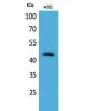
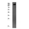
Fig1: Western blot analysis of DSG3 on human DSG3 recombinant protein using anti-DSG3 antibody at 1/1,000 dilution.
Fig2: Western blot analysis of DSG3 on HEK293 (1) and DSG3-hIgGFc transfected HEK293 (2) cell lysate using anti-DSG3 antibody at 1/1,000 dilution.
Fig3: Western blot analysis of DSG3 on A431 cell lysate using anti-DSG3 antibody at 1/1,000 dilution.
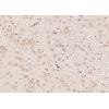
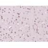
Fig4: Immunohistochemical analysis of paraffin-embedded human esophageal cancer tissue using anti-DSG3 antibody. Counter stained with hematoxylin.
Fig5: Immunohistochemical analysis of paraffin-embedded human esophageal tissue using anti-DSG3 antibody. Counter stained with hematoxylin.
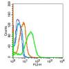
Fig6: Flow cytometric analysis of A431 cells with DSG3 antibody at 1/100 dilution (green) compared with an unlabelled control (cells without incubation with primary antibody; red).
特别提示:本公司的所有产品仅可用于科研实验,严禁用于临床医疗及其他非科研用途!

![Anti-DSG3 antibody [11G-2C]](images/202012/goods_img/92607_G_1607497769393.jpg)