Anti-RF1 antibody
-
概述
- 产品描述Translation is carried out by the ribosome and several associated protein factors through three consecutive steps: initiation, elongation and termination. Termination of protein synthesis takes place when the ribosomal A site is occupied simultaneously by one of three stop codons and by a class 1 translation termination factor. In eukaryotes, this termination factor is the eukaryotic release factor 1 (eRF1), a protein that promotes hydrolysis of the last peptidyl-tRNA on the ribosome. eRF1 activity is stimulated by the association with the GTP-binding protein eRF3. eRF1 forms a quaternary complex with eRF3, GTP and the ribosome. This complex performs a dual role, where, in the "GTP state," it controls the positioning of eRF1 toward the stop codon and peptidyl-tRNA, and, in the "GDP state," it promotes the release of the eRFs from the ribosome. eRF1 contains a highly conserved Asn-Ile-Lys-Ser (NIKS) tetrapeptide, which is essential for the interaction of eRF1 with the ribosome. The gene encoding human eRF1 maps to chromosome 5q31.2.
- 产品名称Anti-RF1 antibody
- 分子量49 kDa
- 种属反应性Human
- 验证应用WB,FC
- 抗体类型小鼠单抗
- 免疫原Recombinant protein
- 偶联Non-conjugated
-
性能
- 形态Liquid
- 浓度2 mg/mL.
- 存放说明Store at +4℃ after thawing. Aliquot store at -20℃ or -80℃. Avoid repeated freeze / thaw cycles.
- 存储缓冲液1*TBS (pH7.4), 1%BSA, 40%Glycerol. Preservative: 0.05% Sodium Azide.
- 亚型IgG1
- 纯化方式Protein A purified.
- 亚细胞定位Cytoplasm.
- 其它名称
- Cl1 protein antibody
- D5S1995 antibody
- ERF antibody
more
-
应用
WB: 1:500-1:2,000
FC: 1:100-1:200
-
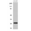
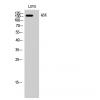
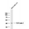
Fig1: Western blot analysis of eRF1 on human eRF1 recombinant protein using anti-eRF1 antibody at 1/1,000 dilution.
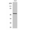
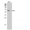
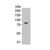
Fig2: Western blot analysis of eRF1 on HEK293 (1) and eRF1-hIgGFc transfected HEK293 (2) cell lysate using anti-eRF1 antibody at 1/1,000 dilution.
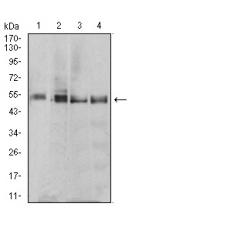
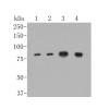
Fig3: Western blot analysis of eRF1 on different cell lysate using anti-eRF1 antibody at 1/1,000 dilution.
Positive control:
Lane 1: MCF-7
Lane 2: T47D
Lane 3: MOLT4
Lane 4: Raji
Fig4: Flow cytometric analysis of Hela cells with eRF1 antibody at 1/100 dilution (green) compared with an unlabelled control (cells without incubation with primary antibody; red).
Fig5: Flow cytometric analysis of HepG2 cells with eRF1 antibody at 1/100 dilution (green) compared with an unlabelled control (cells without incubation with primary antibody; red).
特别提示:本公司的所有产品仅可用于科研实验,严禁用于临床医疗及其他非科研用途!